
Fig. 3
- ID
- ZDB-IMAGE-180622-11
- Publication
- Kague et al., 2018 - Zebrafish sp7 mutants show tooth cycling independent of attachment, eruption and poor differentiation of teeth
- All Figures
- Figures for Kague et al., 2018
|
Fig. 3
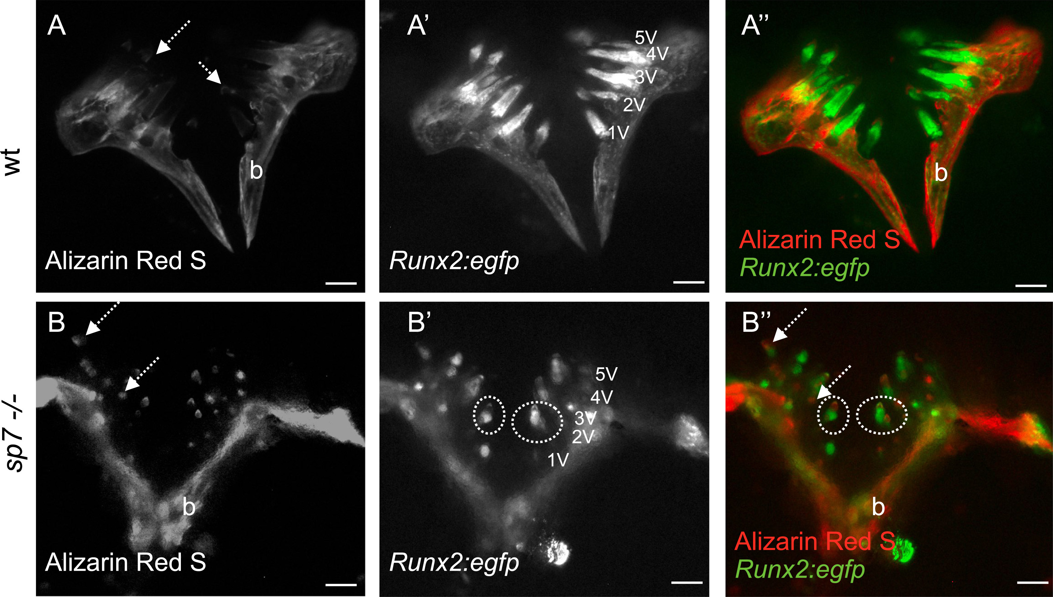
Tooth pattern insp7mutant is unaltered. A-B) wt and sp7 mutant carrying Tg(RUNX2:egfp) and live stained for Alizarin Red S followed by dissection of pharyngeal bone and observed under a stereomicroscope and fluorescent light. Alizarin Red S stains calcium deposition in mineralized tissues. Note tooth tips strongly stained in mutants (dashed arrows) (A,B). Despite the number of teeth in sp7 mutants being seemingly higher, it is still within the range that can be expected: each pharyngeal jaw can show as many as (maximum) 22 teeth (11 functional teeth, 11 replacement teeth). A′, B′) Tg(RUNX2:egfp) showing early odontoblasts and osteoblasts in pulp and bone respectively. Ventral tooth positions are indicated. Some tooth pulps display different shapes (dashed circles). Merged pictures with Alizarin Red S and RUNX2:egfp are shown (A″, B″). Scale bars represent 250 µm.
Reprinted from Developmental Biology, 435(2), Kague, E., Witten, P.E., Soenens, M., Campos, C.L., Lubiana, T., Fisher, S., Hammond, C., Brown, K.R., Passos-Bueno, M.R., Huysseune, A., Zebrafish sp7 mutants show tooth cycling independent of attachment, eruption and poor differentiation of teeth, 176-184, Copyright (2018) with permission from Elsevier. Full text @ Dev. Biol.