
Fig. S4
- ID
- ZDB-IMAGE-180611-58
- Publication
- Freudenblum et al., 2018 - In vivo imaging of emerging endocrine cells reveals a requirement for PI3K-regulated motility in pancreatic islet morphogenesis.
- All Figures
- Figures for Freudenblum et al., 2018
|
Fig. S4
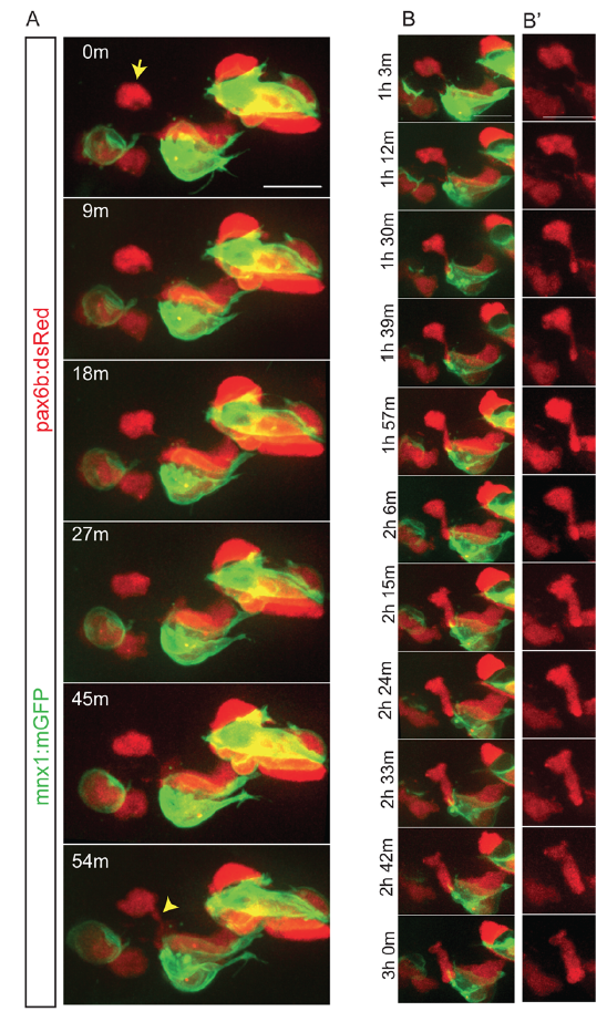
Dynamics of cell coalescence. Related to Figure 3.
Z-projections from time-lapse image series of pancreatic endocrine cells in pax6b:dsRed;mnx1:memGFP transgenics following Notch inhibitor treatment at 4 dpf. Sample was imaged at 7 dpf, with images acquired every 9 minutes. (A) The indicated cell (arrow) forms a connection to a nearby cluster which strengthens over time (bottom frame, arrowhead). (B) Further time points of the sample shown in (A), projections of a subset of zslices to highlight cell shape changes that accompany coalescence of a single cell with a cluster. (B') dsRed channel shown alone for clarity. Scale bars = 10μm.