
Fig. S4
- ID
- ZDB-IMAGE-180608-100
- Publication
- van Boxtel et al., 2017 - Long-Range Signaling Activation and Local Inhibition Separate the Mesoderm and Endoderm Lineages
- All Figures
- Figures for van Boxtel et al., 2017
|
Fig. S4
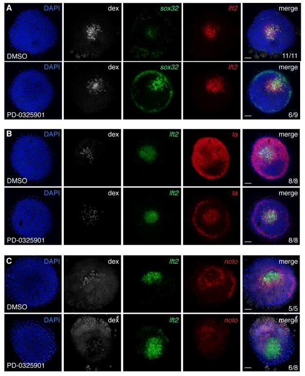
Inhibition of P-Erk signaling promotes the formation of sox32-positive cells in Nodal-expressing clones
(A) Flat-mounted germ ring stage embryos containing a Nodal-expressing clone treated with DMSO or MEK inhibitor PD-0325901 and stained for sox32 and lft2. These are the same embryos as in Figure 4A, except that here individual channels are shown. The number of sox32- positive cells is strongly increased in the PD-0325901-treated embryo.
(B) As in (A), but for lft2 and ta. These are the same embryos as in Figure 4D.
(C) As in (A), but for lft2 and noto. These are the same embryos as in Figure 4E. In (B) and
(C), incubation with PD-0325901 inhibits expression of ta and noto around the clone, confirming that this expression is due to Fgf signaling. All scale bars are 100 μm.
Reprinted from Developmental Cell, 44(2), van Boxtel, A.L., Economou, A.D., Heliot, C., Hill, C.S., Long-Range Signaling Activation and Local Inhibition Separate the Mesoderm and Endoderm Lineages, 179-191.e5, Copyright (2017) with permission from Elsevier. Full text @ Dev. Cell