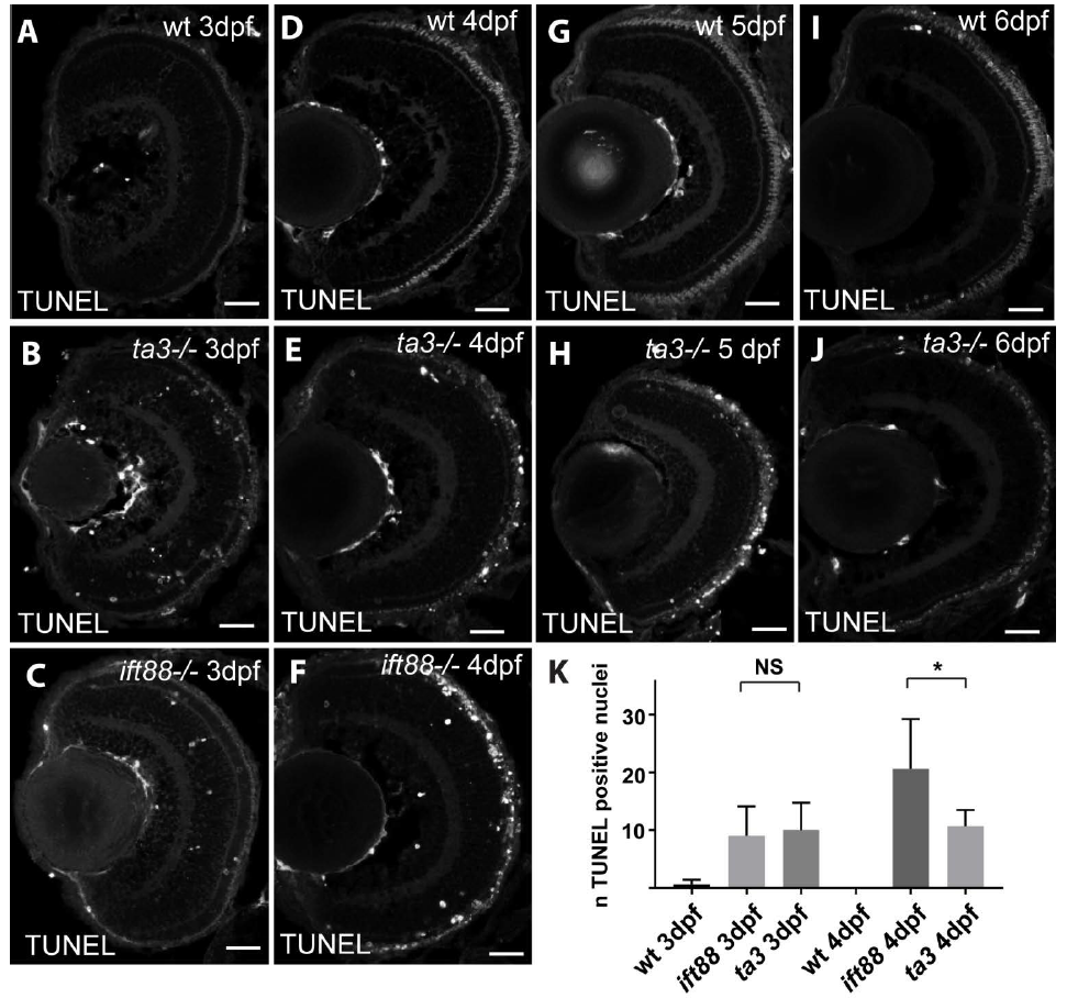
Fig. S4
PR cell death in ta3 mutant retinae TUNEL staining on retinal cryosections at 3 dpf (A-C), 4 dpf (D-F), 5 dpf (G-H) and 6 dpf (I-J), in wildtype (A,D,G and I), talpid3 mutants (B,E,H and J) and ift88 mutants (C and F). Note the strong signal in ift88 mutants at 4 dpf and the comparatively lower amount of cell death in ta3 mutants at the same stage. (K) Quantification of cell death in wt, ift88 and ta3 mutants at 3 and 4dpf. The number of TUNEL-positive nuclei was counted on 5 μm-thick confocal sections of entire retinal sections. Note the stable rate of cell death in ta3 mutants between 3 and 4dpf while ift88 mutants show an increase in cell death at 4dpf compared to 3dpf. Scale bars: 20 μm in all panels.

