
Fig. S16
- ID
- ZDB-IMAGE-180529-12
- Publication
- Sánchez-Iranzo et al., 2018 - Tbx5a lineage tracing shows cardiomyocyte plasticity during zebrafish heart regeneration
- All Figures
- Figures for Sánchez-Iranzo et al., 2018
|
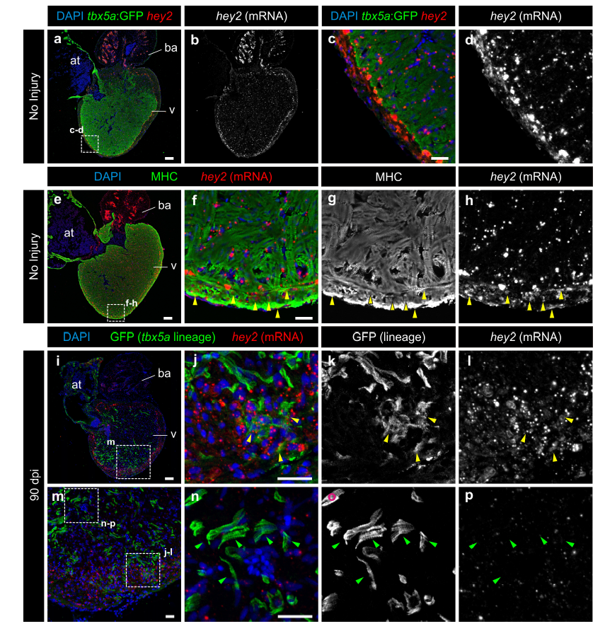
Fig. S16
tbx5a-derived cardiomyocytes within the cortical layer are hey2 positive.
GFP immunofluorescence and hey2 RNAScope in situ detection on sagittal sections of tbx5a:GFP (a–h) or tbx5a:mCherryp-2a-CreERT2;ubb:loxP-lacZ-loxP-GFP double transgenic animals, recombined before injury and fixed at 90 days postinjury (dpi) (i–p). Nuclei are counterstained with DAPI. a–d Uninjured heart. tbx5a:GFP (green) does not co-localize with hey2 (red). Hey2 is expressed at higher and more homogenous levels in the cortical layer. e–h Uninjured heart. anti-Myosin Heavy Chain (MHC) marks cardiomyocytes (green). In the cortical layer, regions of hey2/MHC co-localisation were detected (yellow arrowheads; n=3/4). i–p 90 dpi regenerated hearts. tbx5a:mCherryp-2aCreER T2;ubb:loxP-lacZ-loxP-GFP were treated with two 12 hours pulses of 4-OHT at 6 and 7 days before the injury. GFP+ cells marking the tbx5a lineage within the cortical layer were positive for hey2 (yellow arrowheads, hey2+/GFP+ cells observed in 5 out of 6 hearts. GFP+ cells within the trabecular layer were negative for hey2 (green arrowheads; n=6/6). at, atrium; ba, bulbus arteriosus; v, ventricle. Scale bars, a, e, i 100 μm, c, f, j, n 25 μm