
Fig. 5
- ID
- ZDB-IMAGE-180524-17
- Publication
- Sánchez-Iranzo et al., 2018 - Tbx5a lineage tracing shows cardiomyocyte plasticity during zebrafish heart regeneration
- All Figures
- Figures for Sánchez-Iranzo et al., 2018
|
Fig. 5
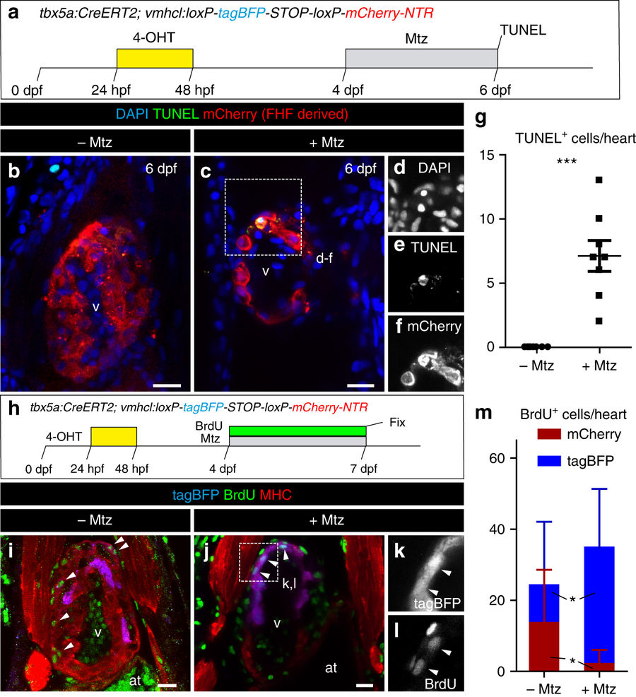
Apoptosis and cell proliferation upon genetic ablation of tbx5a-derived ventricular cardiomyocytes. a tbx5a+ ventricular cardiomyocytes were genetically ablated in tbx5a:CreERT2;vmhcl:loxP-tagBFP-loxP-mCherry-NTR double transgenic zebrafish. Recombination was induced by administration of 4-Hydroxytamoxifen (4-OHT). Cell ablation was induced by administration of Metronidazole (Mtz) from 4 to 7 days postfertilisation (dpf). b, c Optical sections of 6 dpf fish that had been treated with 4-OHT and Mtz as indicated in a (b) or only with 4-OHT c immunostained for mCherry (red) and terminal deoxynucleotidyl transferase (TdT)-mediated dUTP nick end labeling (TUNEL) (green). Note that some rounded mCherry+ cells are TUNEL+. d–f Single channels of selected area in c. g Quantification of the number of TUNEL+ trabecular and cortical mCherry+ cardiomyocytes per heart from animals of the +Mtz (n = 8) and –Mtz (n = 8) group, mean ± SD; ***P = 0.0004 by Mann–Whitney non-parametric t-test. h Schematic representation of the 5-Bromo-2´-deoxyuridine (BrdU) treatment to assess proliferation. i, j Fish were treated with 4-OHT and BrdU i or with 4-OHT, Mtz, and BrdU j. k, l Single channels of the boxed area in j. m Quantification of BrdU+/mCherry+ and BrdU+/tagBFP+ cells per heart. Shown are means ± SD (n = 8 for − Mtz hearts and n = 11 for + Mtz hearts, from two separate independent experiments) *P = 0.0240 for tagBFP+ cells and P = 0.0371 for mCherry+ cells by two-tailed t-test. at, atrium; hpf, hours postfertilization; v, ventricle. Scale bars, 25 μm