
Fig. 4
- ID
- ZDB-IMAGE-180524-16
- Publication
- Sánchez-Iranzo et al., 2018 - Tbx5a lineage tracing shows cardiomyocyte plasticity during zebrafish heart regeneration
- All Figures
- Figures for Sánchez-Iranzo et al., 2018
|
Fig. 4
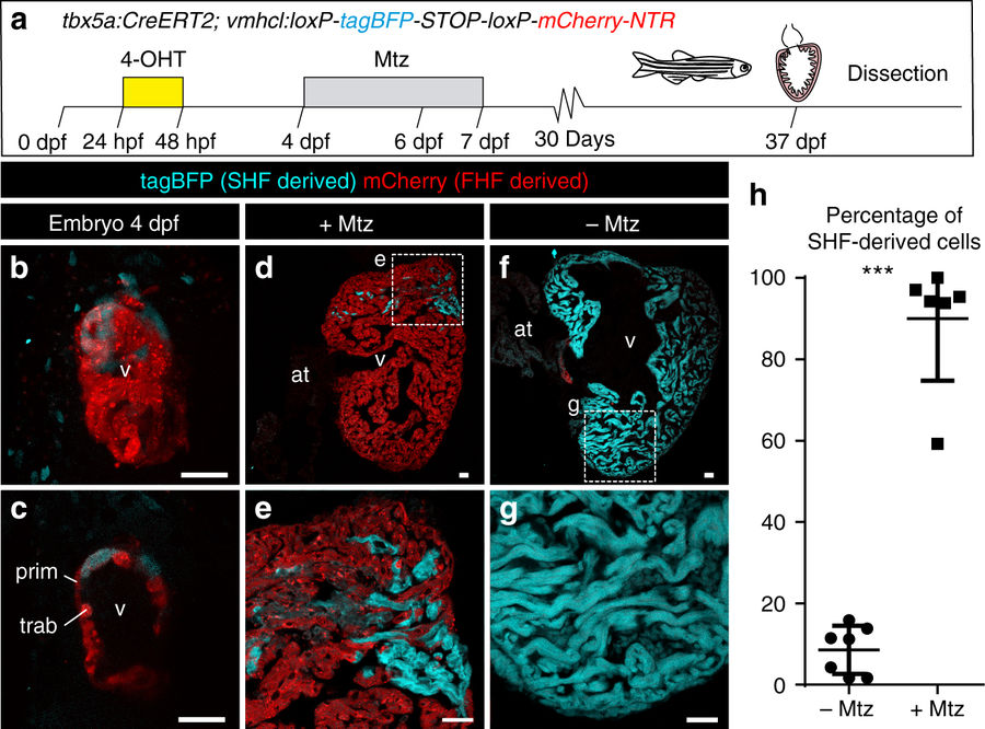
Genetic ablation of tbx5a-derived ventricular cardiomyocytes. a tbx5a+ ventricular cardiomyocytes were genetically ablated in tbx5a:CreERT2;vmhcl:loxP-tagBFP-loxP-mCherry-NTR double transgenic zebrafish. Recombination was induced by administration of 4-Hydroxytamoxifen (4-OHT). Cell ablation was induced by administration of Metronidazole (Mtz) from 4 to 7 days postfertilisation. b, c Ventral views of larval hearts at 4 dpf (maximal projection and optical section, respectively). Anterior is to the top. The proximal ventricle, including primordial layer and trabeculae, is completely mCherry+ and the distal ventricle is blue (tagBFP+); n = 7/7. d, e Sagittal section of the ventricle of an adult recombined heart. Most cells are mCherry+. Only the tbx5a− region is tagBFP. n = 7/7. f, g, Sagittal section of a Mtz-treated fish. Most of the cardiomyocytes are tagBFP+; n = 6/6. h, Quantification of the percentage of myocardium that is tagBFP+ (SHF-derived), mean±SD; *** P < 0.0001 by two-tailed unpaired t-test. at, atrium; prim, primordial layer; SHF, second heart field; trab, trabeculae; v, ventricle. Scale bars, 25 μm