
Fig. 2
- ID
- ZDB-IMAGE-180516-13
- Antibodies
- Publication
- Ojeda Naharros et al., 2017 - Loss-of-function of the ciliopathy protein Cc2d2a disorganizes the vesicle fusion machinery at the periciliary membrane and indirectly affects Rab8-trafficking in zebrafish photoreceptors
- All Figures
- Figures for Ojeda Naharros et al., 2017
|
Fig. 2
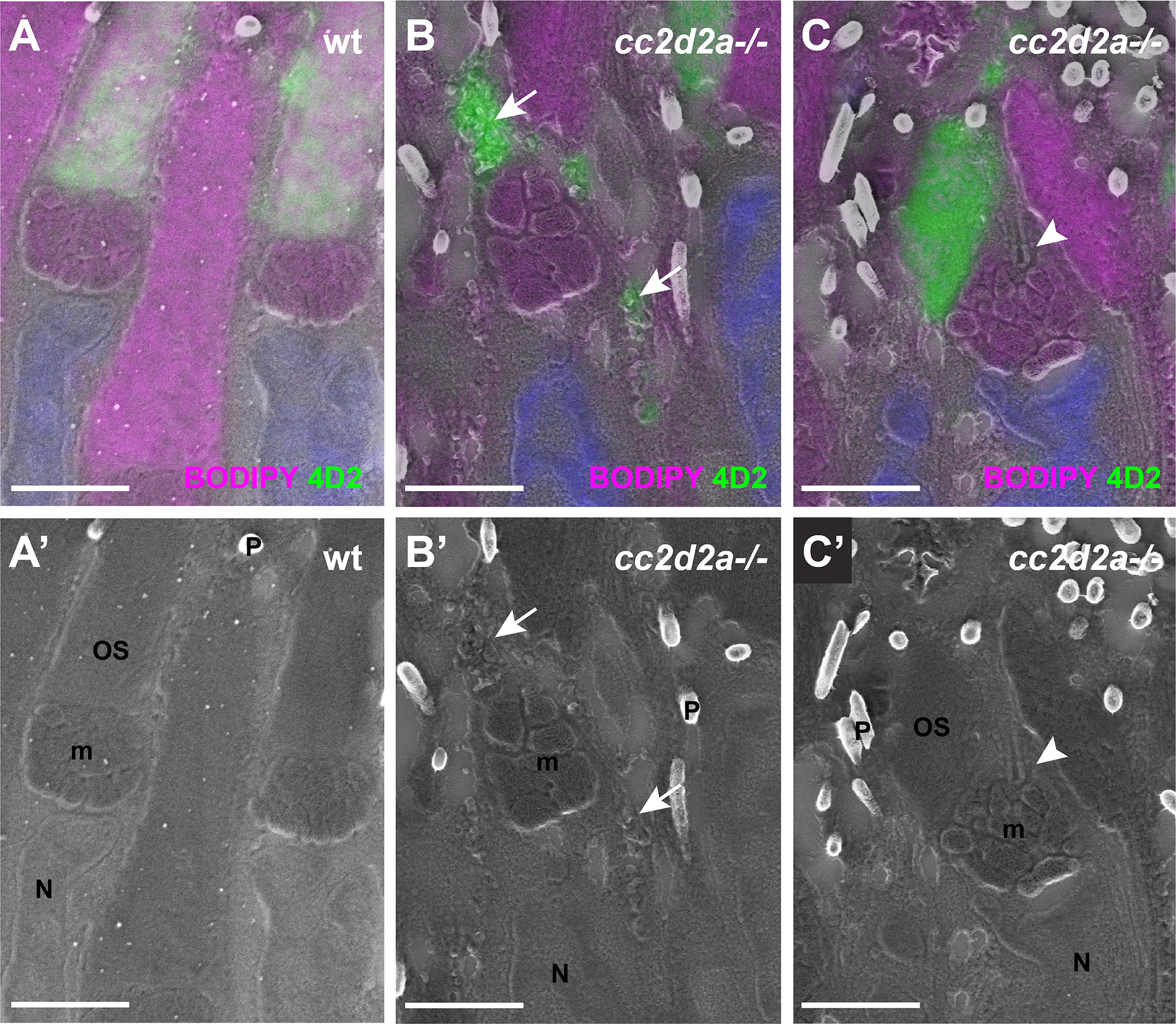
Accumulated vesicles in cc2d2a-/- PRs are opsin-carrier-vesicles (A-C) 5 dpf correlative light and electron microscopy (CLEM) image of retinal sections stained with BODIPY (magenta) to mark membranes of the outer segment and the mitochondrial cluster and with 4D2 (green) to label rhodopsin and red-green cone opsin. (A’-C’) are the corresponding scanning electron microscopy (SEM) images. Note that while 4D2 staining only localizes at the outer segments of wild-type (wt) PRs (A), it is visible in accumulated vesicles in cc2d2a-/- PRs (B, arrows) and in dysmorphic outer segments (C). Also note normal cilium docking in cc2d2a-/- PRs (C-C’, arrowhead). Scale bars are 2 μm in all panels. m mitochondria, N nuclei, OS outer segments, P pigment in melanosomes, wt wild-type.