
Image
|
Figure Caption
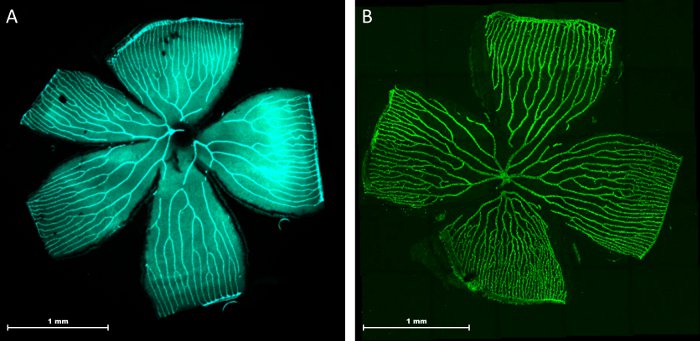
Fig. 9
Representative pictures of adult tg(fli:EGFP) zebrafish retinal vasculature. Two typical morphological examples of the retinal vasculature are shown: The central optic artery spreads into 5-7 main vessels, which then branch into a succession of arcades. All further vessels drain into the circumferential vein (CV) limiting the outer part of each petal. Visualization via fluorescence microscopy at 2.5x magnification (A). Visualization of the retinal vasculature by confocal laser scanning microscopy through a combined 5x5 single image tile scan (B).
Acknowledgments
This image is the copyrighted work of the attributed author or publisher, and
ZFIN has permission only to display this image to its users.
Additional permissions should be obtained from the applicable author or publisher of the image.
Full text @ J. Vis. Exp.