
Image
|
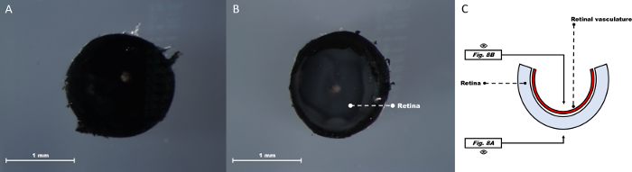
Figure Caption
Fig. 8
Adult zebrafish eye after removal of RPE/choroid and truncated optic nerve. Optic nerve side view shows the retina with a truncated optic nerve (A). Corneal side view with a direct look onto the most inner layer of the retina (B). Schematic depiction of the zebrafish eye at this step (C).
Acknowledgments
This image is the copyrighted work of the attributed author or publisher, and
ZFIN has permission only to display this image to its users.
Additional permissions should be obtained from the applicable author or publisher of the image.
Full text @ J. Vis. Exp.