
Image
|
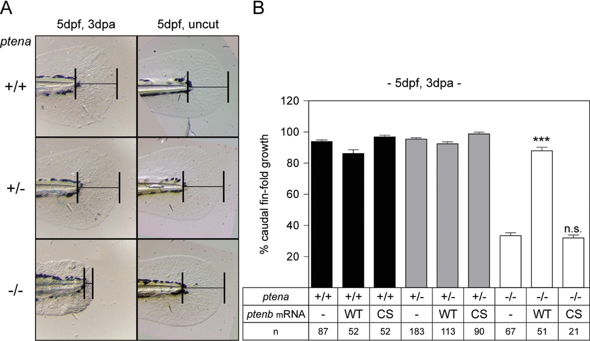
Figure Caption
Fig. 1
Impaired caudal fin‐fold regeneration in Pten deficient embryos. (A) Embryos from a ptena+/−ptenb−/− in‐cross were micro‐injected at the one‐cell stage with synthetic mRNA encoding WT Ptenb (WT), catalytically inactive Ptenb‐C124S (CS), or were not injected (−). At 2 dpf the caudal fin‐fold was amputated and regeneration was assessed at 3 dpa (i.e., 5 dpf, 3 dpa); equivalent uncut controls were included (i.e., 5 dpf, uncut). All embryos were genotyped. Representative images of non‐injected embryo caudal fin‐folds are shown, and of WT Ptenb or Ptenb‐C124S‐injected embryos in Fig. S1. (B) The means of caudal fin‐fold growth following amputation are depicted relative to caudal fin‐fold growth of uncut ptena+/+ptenb−/− controls. Means of micro‐injected amputated ptena−/−ptenb−/− embryos were compared to non‐injected amputated ptena−/−ptenb−/− embryos. Significance: ***p < 0.001; n.s., not significant; error bars indicate standard error of the mean. Data pooled from multiple experiments
Acknowledgments
This image is the copyrighted work of the attributed author or publisher, and
ZFIN has permission only to display this image to its users.
Additional permissions should be obtained from the applicable author or publisher of the image.
Full text @ Regeneration (Oxf)