
Image
|
Figure Caption
Fig. 3
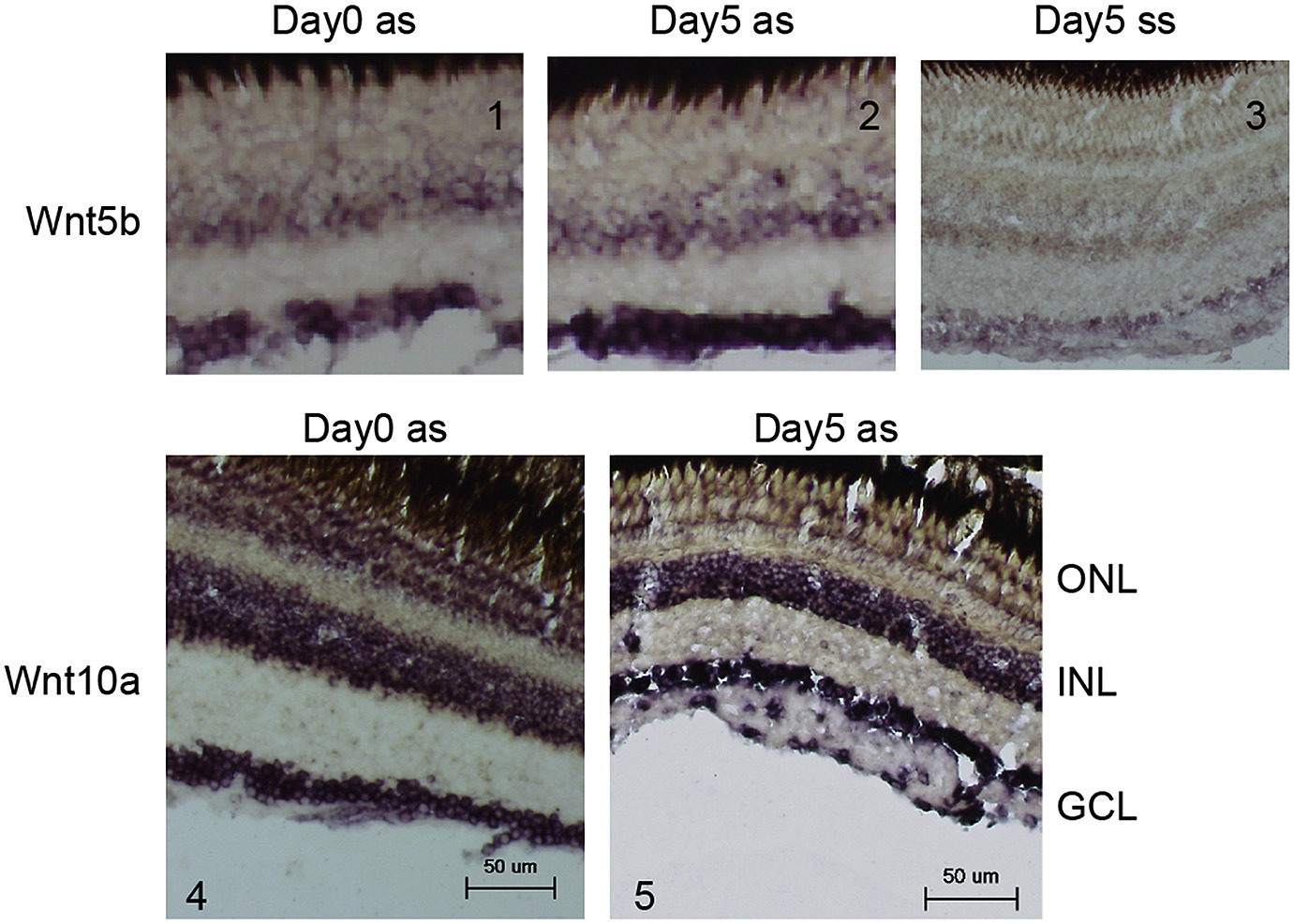
Cellular localization of Wnt5b and Wnt10a in the retina. Zebrafish retinas were isolated at day 0 (panels 1 and 4) or day 5 (panels 2, 3 and 5). Each slice was hybridized with Wnt5b anti-sense cRNA (panels 1 and 2), Wnt5b sense cRNA (panel 3) or Wnt10a anti-sense cRNA (panels 4 and 5). ONL: outer nuclear layer; INL: inner nuclear layer; GCL: ganglion cell layer. Scale bar: 50 μm.
Acknowledgments
This image is the copyrighted work of the attributed author or publisher, and
ZFIN has permission only to display this image to its users.
Additional permissions should be obtained from the applicable author or publisher of the image.
Full text @ Neurochem. Int.