
Fig. 3-S1
- ID
- ZDB-IMAGE-180508-13
- Publication
- Aguillon et al., 2018 - Cell-type heterogeneity in the early zebrafish olfactory epithelium is generated from progenitors within preplacodal ectoderm
- All Figures
- Figures for Aguillon et al., 2018
|
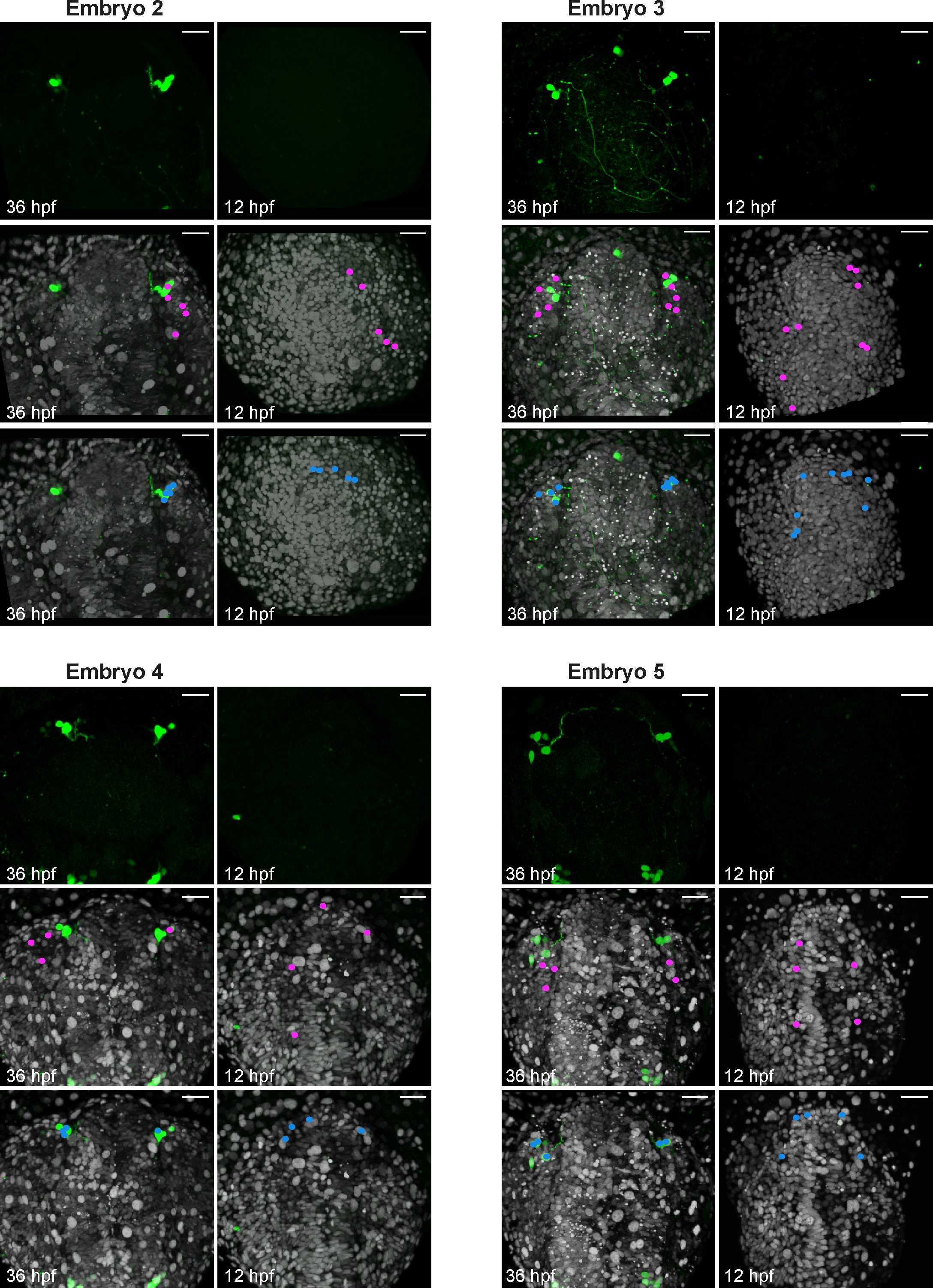
Fig. 3-S1
Backtracking data from individual Tg(gnrh3:eGFP) embryos.
Confocal projections extracted from 4D datasets at 36 and 12 hpf for 4 embryos analysed and not shown in Figure 3 showing the GFP channel alone, and the position of the backtracked nuclei of gnrh3:eGFP-negative (pink) and gnrh3:eGFP-positive (blue) cells at both timepoints. Backtracked nuclei for the lens were only performed for Embryo 1 and are not shown. Embryos are shown with anterior up; whereas the GFP expression detected caudally is ectopic and does not reflect bona fide gnrh3 expression, the GFP+ axons seen in Embryo 3 extend from the trigeminal ganglia, which do expression gnrh3 endogenously. Scalebars represent 40 μm.