
Fig. 4-S1
- ID
- ZDB-IMAGE-180504-3
- Publication
- Montague et al., 2017 - Vg1-Nodal heterodimers are the endogenous inducers of mesendoderm
- All Figures
- Figures for Montague et al., 2017
|
Fig. 4-S1
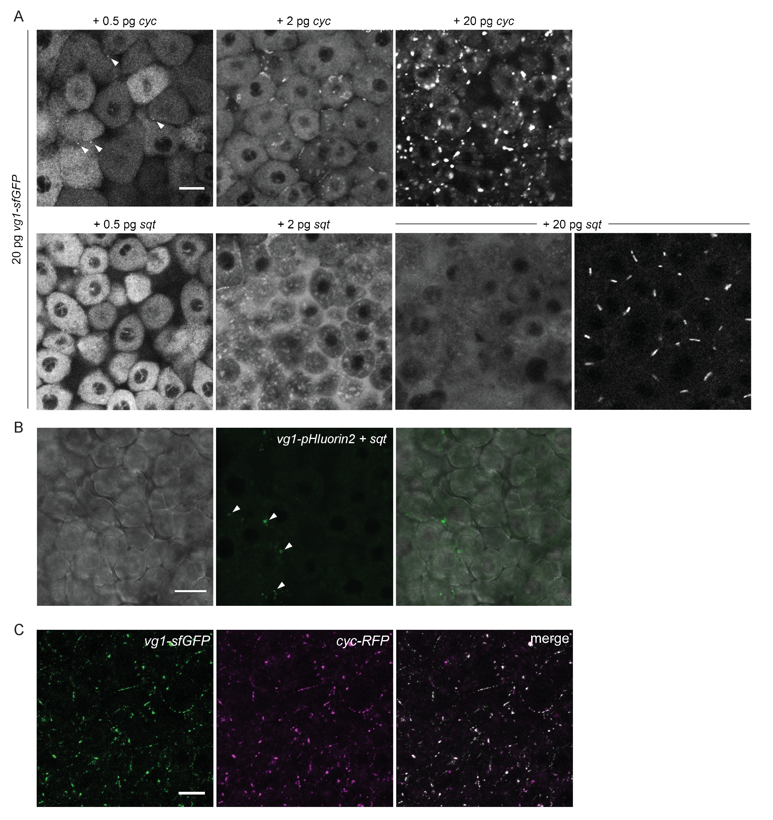
Vg1 secretion requires Nodal.
(A) Mvg1 embryos co-injected with 20 pg of vg1-sfGFP mRNA and 0.5 pg, 2 pg or 20 pg of cyc or sqt mRNA. These images were all captured under the same microscope settings. Arrowheads indicate small extracellular puncta. Note that the puncta increase in size with increasing concentrations of cyc mRNA. Co-expression of vg1-sfGFP mRNA and sqt mRNA can result in extracellular diffuse signal and/or extracellular puncta. Scale bar, 17 um. See Table 1 for quantification. (B) Mvg1 embryo co-injected with pH-sensitive fluorescent vg1 (vg1-pHluorin2) and sqt. Arrowheads indicate fluorescent puncta. Scale bar, 17 um. (C) Z-stack of Mvg1 embryo co-injected with 50 pg of vg1-sfGFP and 50 pg of cyc-sfGFP. Scale bar, 17 um.