Image
Figure Caption
Fig. 1
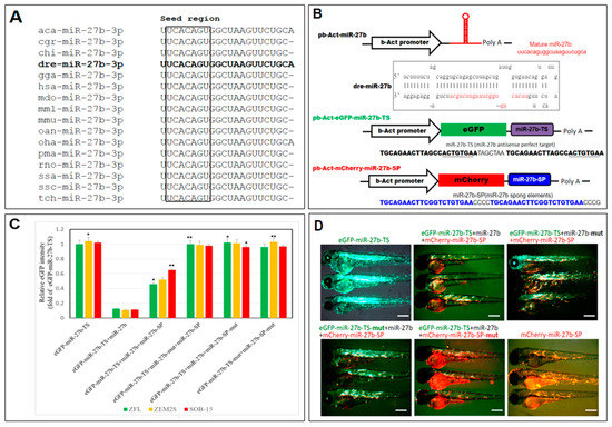
Design and validation of a miR-27b-sponge (miR27b-SP). (A) Alignment of the mature miR-27b sequence is perfectly conserved across many species, including arboreal lizard (Anolis carolinensis; aca-miR-27b), chinese hamster (Cricetulus griseus; cgr-miR-27b), domestic goat (Capra hircus; chi-miR-27b), zebrafish (Danio rerio; dre-miR-27b), red junglefowl (Gallus gallus; gga-miR-27b), humans (Homo sapiens; hsa-miR-27b), gray short-tailed opossum (Monodelphis domestica; mdo-miR-27b), rhesus macaque (Macaca mulatta; mml-miR-27b), mouse (Mus musculus; mmu-miR-27b), platypus (Ornithorhynchus anatinus; oan-miR-27b), king cobra (Ophiophagus hannah; oha-miR-27b), sea lamprey (Petromyzon marinus; pma-miR-27b), rat (Rattus norvegicus; rno-miR-27b), Atlantic salmon (Salmo salar; ssa-miR-27b), wild boar (Sus scrofa; ssc-miR-27b) and chinese tree shrew (Tupaia chinensis; tch-miR-27b). (B) Cloning of pri-miR-27b and miR27b-SP into b-Act expression vectors. Stem-loop structure of premiR-27b is shown, in which mature miR-27b is highlighted in red. (C) In vitro EGFP reporter assays were performed to confirm the direct interaction between miR-27b and the target sequences. ZFL, ZEM2S, and SOB-15 cells were transfected with indicated b-Act-miR-27b plasmids, and the EGFP intensity was measured. * p < 0.01, and ** p < 0.005. (D) In vivo EGFP reporter assays were performed to confirm the direct interaction between miR-27b and the target sequences in six days post fertilization (dpf) zebrafish larvae.
Acknowledgments
This image is the copyrighted work of the attributed author or publisher, and
ZFIN has permission only to display this image to its users.
Additional permissions should be obtained from the applicable author or publisher of the image.
Full text @ Int. J. Mol. Sci.

