
IMAGE
Fig. S2
- ID
- ZDB-IMAGE-180502-6
- Publication
- Li et al., 2017 - Axonemal dynein assembly requires the R2TP complex component Pontin
- All Figures
- Figures for Li et al., 2017
Image
|
Figure Caption
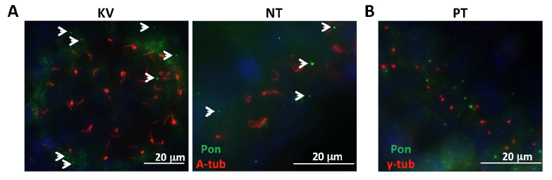
Fig. S2
Subcellular localization of Pontin in zebrafish embryos. (A) Pontin distribution in the Kupffer’s vesicle (KV, 8-somite stage) and neural tube (NT, 24 hpf). Cilia are labeled with anti-acetylated-tubulin (A-Tub) in red and Pontin is labeled with anti-Pontin (Pon) in green. White arrowheads point to Pontin puncta. (B) Pontin puncta are not associated with the basal body marker anti-γ-tubulin in the pronephric tubule (PT) at 24 hpf. Pontin is labeled with anti-Pontin (Pon) in green and basal bodies are labeled with anti-γ-tubulin (γ-tub) in red. Nuclei are labeled with DAPI in blue.
Acknowledgments
This image is the copyrighted work of the attributed author or publisher, and
ZFIN has permission only to display this image to its users.
Additional permissions should be obtained from the applicable author or publisher of the image.
Full text @ Development