
|
Fig. 5
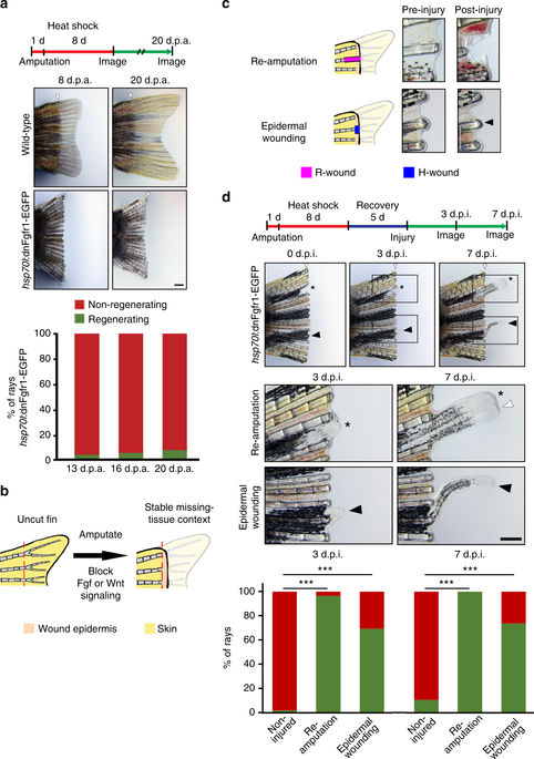
Epidermal wounds inflicted on dormant zebrafish fins induce regeneration. a Transient overexpression of a dominant-negative Fgf receptor-1 (dnFgfr1-GFP) until 8 days post-amputation (d.p.a.) blocked fin regeneration, which spontaneously resumed only in a minority of fin rays after relief from transgene expression. White arrowheads mark the amputation plane, n = 9 fish, 163 rays. b A stable missing-tissue context (dormant fin) can be produced by transient blockage of Fgf or Wnt/β−catenin signaling, which allows for wound epidermis formation but stably blocks fin ray regeneration. c Injury regimes applied to dormant fins. Re-amputation represents an R-wound, epidermal wounding an H-wound. Arrowhead points at epidermal wound. d Re-amputation (asterisk), as well as epidermal wounding (black arrowhead) re-initiated regenerative growth. Lower panels show magnification of boxed areas. The graph depicts the fraction of non-injured, re-amputated and epidermally wounded rays displaying regenerative growth at 3 and 7 days post-injury (d.p.i.). ***p < 0.001 χ 2 test. n (non-injured) = 449 rays, 61 fish, 3 experiments; n (re-amputated) = 30 rays, 28 fish, 1 experiment, n (epidermally wounded) = 84 rays, 61 fish, 3 experiments. Note that uninjured rays located next to regenerating rays frequently exhibited bystander growth (white arrowhead in box) in both wounding scenarios (see also Supplementary Fig. 9). Scale bars: 500 μm