
Fig. 2
- ID
- ZDB-IMAGE-180502-13
- Publication
- Barsh et al., 2017 - Vagus Motor Neuron Topographic Map Determined by Parallel Mechanisms of hox5 Expression and Time of Axon Initiation
- All Figures
- Figures for Barsh et al., 2017
|
Fig. 2
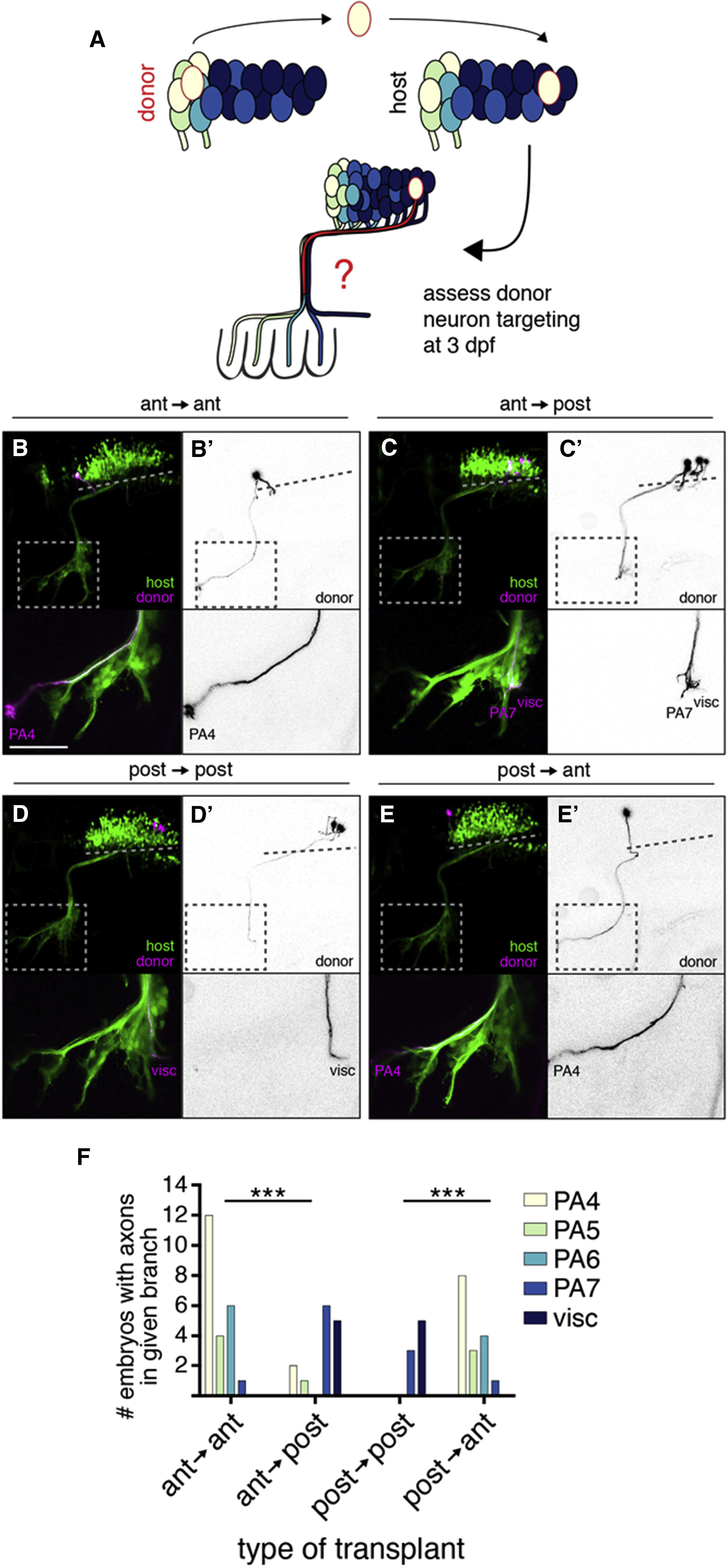
Vagus Motor Neuron Position Determines Axon Target
(A) Schematic of postmitotic neuron transplantation approach. mX neurons are transplanted homotopically or heterotopically (pictured) before axon formation, and donor axon targeting is assayed at 3 dpf.
(B–E) Examples of homotopic (B and D) and heterotopic (C and E) transplants. Donor-derived neurons are marked by Tg(isl1:Kaede) in magenta and black on white (B′–E′). Host motor neurons are marked by Tg(isl1:GFP) in green. Dotted lines indicate length of mX territory; dotted boxes indicate region shown in lower panels.
(F) Quantification of transplant results showing number of host embryos with a donor axon in a given branch. Statistical analysis was done with Fisher’s exact test (see STAR Methods for details). Ant → ant: n = 14 host embryos; ant → post: n = 12; post → post: n = 7; post → ant: n = 12.