Fig. S2
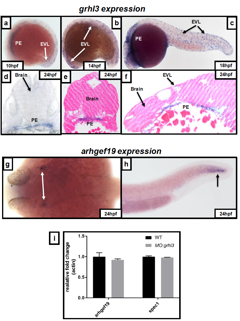
Expression patterns of grhl3 and arhgef19 in early zebrafish development.
(A-F) grhl3 is expressed within the pharyngeal endoderm (PE) from 10 hpf (A), a timepoint at which expression within the EVL is also visible. Expression within both these regions persists until at least 18 hpf (B-C). grhl3 expression is not seen in brain; non-specific probe trapping (C) is confirmed as artefact following both brightfield imaging of coronal sections (D) and subsequent H&E staining of both coronal (E) and sagittal (F) sections. Note persistent expression of grhl3 within the PE (D-F) and occasional puncta of staining within the EVL (F). (G-H) Expression of arhgef19 is seen in the EVL, lateral to the MHB (arrows, G), as well as in the EVL at the posterior-most region of the extending tail (arrow, H) at 24 hpf. (I) Q-RT-PCR showing expected non-significant decrease in arhgef19 and spec1 transcripts in MO:grhl3 injected embryos.

