
IMAGE
Fig. 2
- ID
- ZDB-IMAGE-180425-24
- Publication
- Nagata et al., 2017 - Heart Failure Phenotypes Induced by Knockdown of DAPIT in Zebrafish: A New Insight into Mechanism of Dilated Cardiomyopathy
- All Figures
- Figures for Nagata et al., 2017
Image
|
Figure Caption
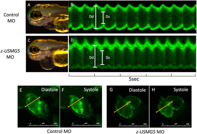
Fig. 2
Representative images of zebrafish hearts at 72 hpf. Brightfield and fluorescent microscopy images of zebrafish embryos injected control MO (A,B) and z-usmg5 MO (C,D), respectively. z-usmg5 MO injected embryos showed swollen pericardial sacs (C) and reduced ventricular contraction (D,G,H) compared to the control MO embryos (B,E,F). hpf: hours post fertilization, MO: morpholino oligonucleotide, Dd: diastolic diameter, Ds: systolic diameter.
Figure Data
Acknowledgments
This image is the copyrighted work of the attributed author or publisher, and
ZFIN has permission only to display this image to its users.
Additional permissions should be obtained from the applicable author or publisher of the image.
Full text @ Sci. Rep.