
|
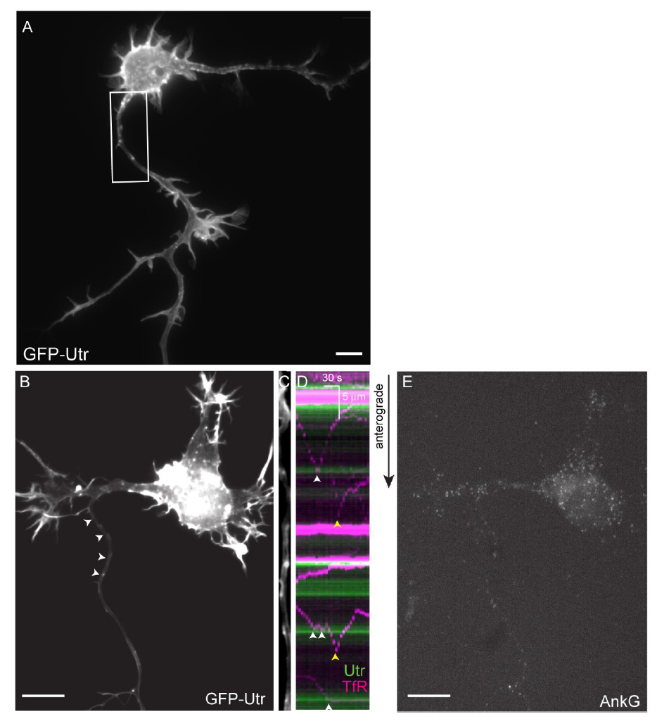
Fig. S2
Vesicles carrying a dendritic protein tend to halt and reverse at sites near actin patches, related to Fig. 1. (A) Low magnification image of neuron whose proximal axon is shown in Fig. 1(E) (B) 3 DIV cortical neuron co-expressing GFP-Utr and TfR-mCherry (not shown). Arrowheads point to axon. (C) Straightened axon from neuron in (B). (D) Kymograph of neuron in (B, C) showing GFP-Utr (green) and TfR-mCherry (purple). White arrowheads indicate places where TfR containing vesicles halted near actin patches, yellow arrowheads indicate places where halting took place away from patches. (E) Ankyrin G staining of neuron in (B-D) showing a lack of Ankyrin G expression. Scale bar 10 μm.