
Fig. 6
- ID
- ZDB-IMAGE-180424-23
- Publication
- Uribe et al., 2017 - Retinoic acid temporally orchestrates colonization of the gut by vagal neural crest cells
- All Figures
- Figures for Uribe et al., 2017
|
Fig. 6
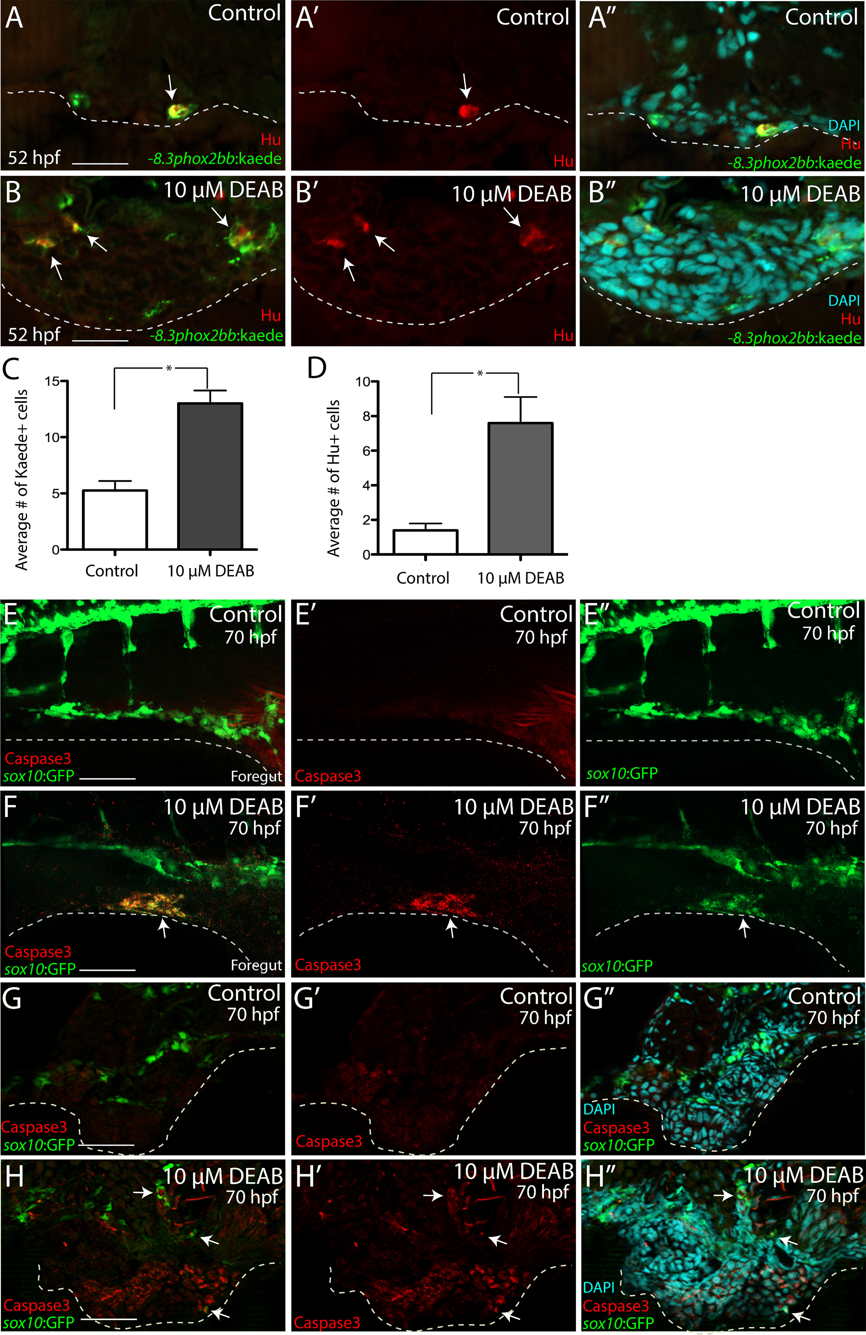
RA depletion causes accumulation of enteric progenitors in the ventral mesenchyme near the foregut and their apoptosis. (A-A’’-B-B’’) At 52 hpf, transverse sections through the foregut of control (A-A’’) and DEAB treated larvae (B-B’’) reveal the location of -8.3phox2bbb:Kaede+/Hu+ enteric progenitors (arrows), (C-D) Bar graphs depict the average number of Kaede+ enteric progenitors (C) and Hu+ enteric neurons (D) in control and DEAB treated larval foregut sections. n=5 embryos for each condition. Error bars indicate +/- S.E.M. *, p<.05 with Student's t-test. (E-E’’-F-F’’) Whole mount double immunochemistry against activated-Caspase3 (red) and GFP (green) in sox10:GFP control (E-E’”) and DEAB treated (F-F’’) larval fish. GFP+/Caspase3+ cells are present along the foregut of DEAB treated fish, however not detected in control embryos. (G-G’’, H-H’’) At 70 hpf, transverse sections through the foregut of control (G-G’’) and DEAB treated larvae (H-H’’) show the location of sox10:GFP+ and Caspase3+ cells (red), which reveals neural crest cells that are Caspase3+ surrounding the gut in DEAB treated fish, when compared to controls. Scale bar in A, B, G-H: 20 μM; scale bar in E,F: 60 μM.
Reprinted from Developmental Biology, 433(1), Uribe, R.A., Hong, S.S., Bronner, M.E., Retinoic acid temporally orchestrates colonization of the gut by vagal neural crest cells, 17-32, Copyright (2017) with permission from Elsevier. Full text @ Dev. Biol.