
Fig. 2
- ID
- ZDB-IMAGE-180424-19
- Publication
- Uribe et al., 2017 - Retinoic acid temporally orchestrates colonization of the gut by vagal neural crest cells
- All Figures
- Figures for Uribe et al., 2017
|
Fig. 2
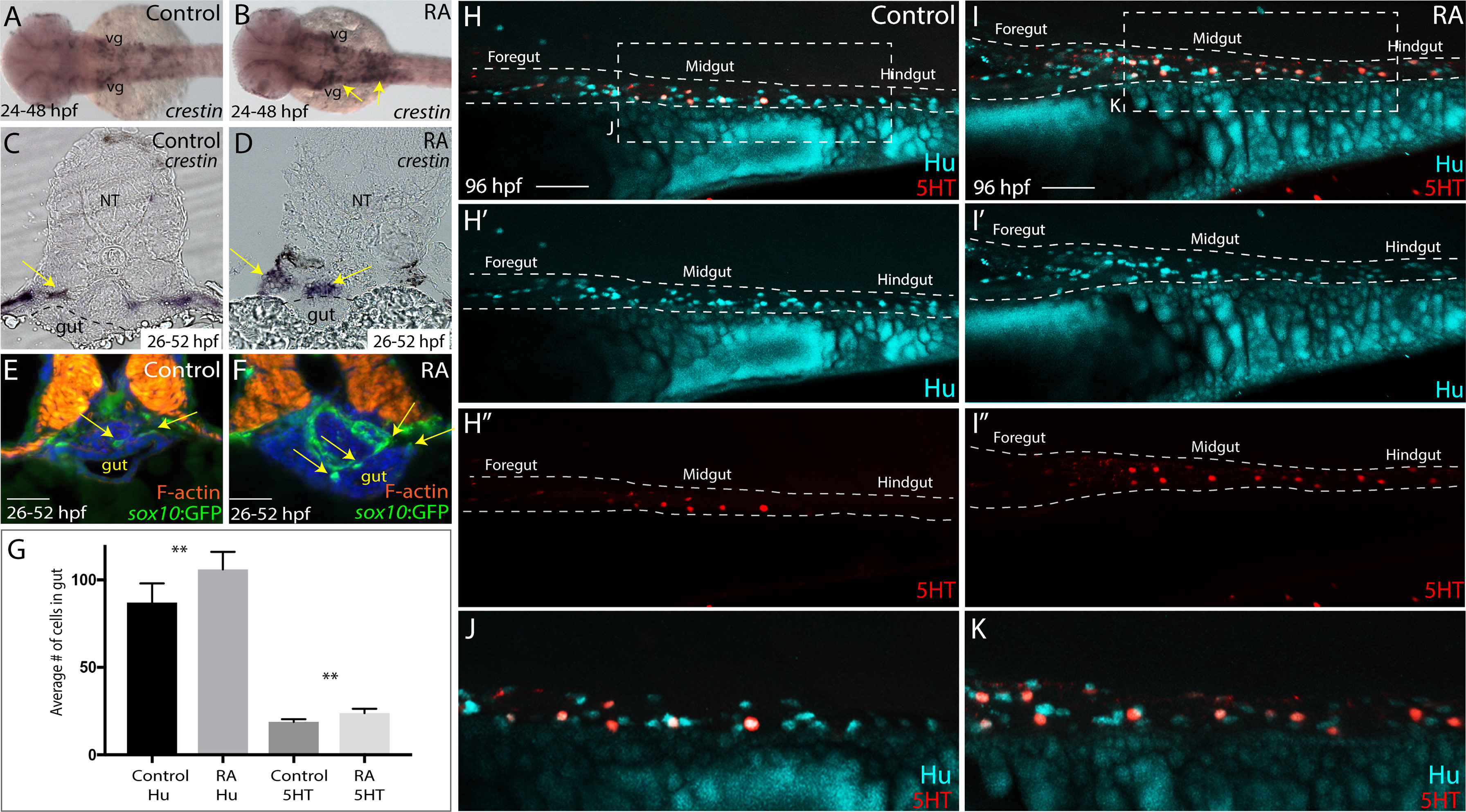
RA treatment during enteric neural crest migration stages enhances enteric neuron numbers. (A-B) A dorsal view following whole-mount in situ hybridization at 48 hpf for crestin in control (A) and RA-treated larvae (B) reveals neural crest localization along the embryo. RA-treated embryos display an expansion of neural crest, arrows (B), when compared to control (A). vg-vagal (C-D) Cryosections through the foregut of larvae at 52 hpf following in situ hybridization for crestin in (C) control and (D) RA-treated larvae shows an expanded distribution of crestin+ neural crest near the vicinity of the gut (arrows), when compared with control larvae. NT- neural tube (E-F) Transverse sections depicting the localization of sox10:GFP+ neural crest in (E) control and (F) RA-treated larvae shows an expansion of neural crest near the vicinity of the gut (arrows), when compared with control larvae. Scale bars, 50 μM (G) Bar graph to represent the average number of Hu+ and 5HT+ neurons along the gut in control and RA-treated larvae at 96 hpf. Error bars indicate +/- S.E.M. , n = 6 embryos for each condition. (H-I) Lateral views of the gut at 96 hpf following whole-mount antibody staining to detect Hu+/5HT+ enteric neurons in (H-H’’) control and (I-I’’) RA-treated larvae. (J) Zoomed in view of control larval fish depicted in H. (K) Zoomed in view of RA-treated larval fish depicted in I. Scale bars, 50 μM.
Reprinted from Developmental Biology, 433(1), Uribe, R.A., Hong, S.S., Bronner, M.E., Retinoic acid temporally orchestrates colonization of the gut by vagal neural crest cells, 17-32, Copyright (2017) with permission from Elsevier. Full text @ Dev. Biol.