
Fig. S20
- ID
- ZDB-IMAGE-180420-52
- Publication
- Richter et al., 2017 - Small molecule screen in embryonic zebrafish using modular variations to target segmentation
- All Figures
- Figures for Richter et al., 2017
|
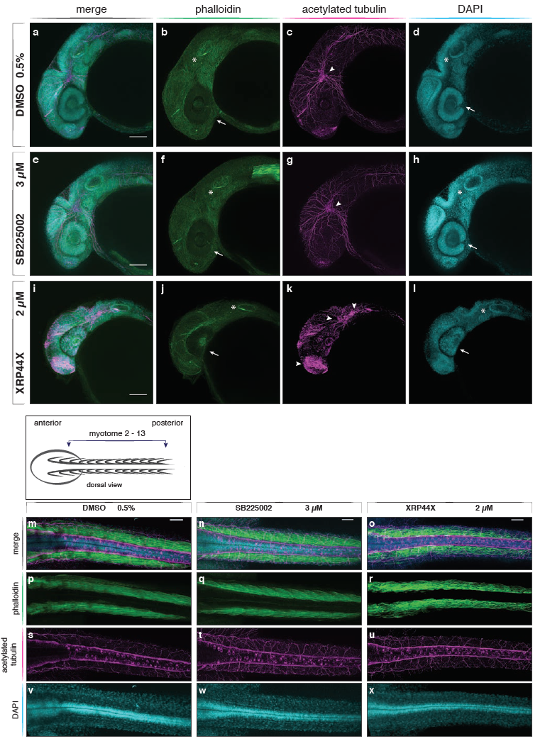
Fig. S20
Head structures, neurites and dorsal view of trunk in SB225002 and XRP44X treated embryos
Confocal images of DMSO control and small molecule treated embryos at 36 hpf. F-actin visualized with phalloidin (green), acetylated tubulin by immunostaining (magenta) and nuclei with DAPI (cyan). (a-l) Maximum projection of a stack of confocal sections of the lateral head of DMSO control and small molecule treated embryos. Scale bar is 100 μm. Control (a-d) and SB225002 (e-h) treated embryos show comparable development of main organs and tissue of the head: eyes with lens and retina (arrows), otic vesicles (asterisks) and neurons of the central nervous system (arrowheads). XRP44X treatment (i-l) led to a reduced eye size and loss of the ventral fissure (j), abnormal distribution of acetylated tubulin and accumulation in the head, developing mouth and olfactory organs (asterisks, k). (m-x) Maximum projection of a stack of confocal sections of the dorsal trunk area of DMSO control and small molecule treated embryos. Scale bar is 50 μm. Individual myotome structure is visible in DMSO and SB225002 treated embryos (p,q), but is less organised in XRP44X treatments (r) where individual myotomes are not detectable. (s-u) Neurons of the peripheral nervous system developed all along the anterior posterior axis in control embryos and all treatments. (v-x) Nuclei along the axis show a comparable distribution of cells between control and drug treatments.