
Fig. 4
- ID
- ZDB-IMAGE-180420-42
- Publication
- Richter et al., 2017 - Small molecule screen in embryonic zebrafish using modular variations to target segmentation
- All Figures
- Figures for Richter et al., 2017
|
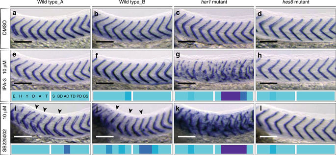
Fig. 4
Mutant background changed sensitivity to small molecule perturbation. Wild type, her1 and hes6 mutant embryos at 36 hpf after small molecule treatment as indicated, xirp2a expression in myotome boundaries shown for segments 5–15. a–d Myotome boundaries of each genotype normal in 0.5% DMSO control. 10 µM IPA-3 normal segmentation in wild type replicates e, f and in the hes6 mutant h. g 10 µM IPA-3 in her1 mutants, severe myotome boundary disruptions in trunk. i, j 10 µM SB225002 in wild type, mild myotome boundary defects of boundaries 4–10 (arrowheads). k 10 µM SB225002 in her1 mutant, severe boundary disruptions, defects extended posteriorly along axis. l 10 µM SB225002 in hes6 mutant, no effect on myotome boundary. Embryos had otherwise normal morphology, as indicated by phenotypic vector fingerprints. Scale bar: 100 µm