
Fig. S4
- ID
- ZDB-IMAGE-180420-37
- Publication
- Dogra et al., 2017 - Opposite effects of Activin type 2 receptor ligands on cardiomyocyte proliferation during development and repair
- All Figures
- Figures for Dogra et al., 2017
|
Fig. S4
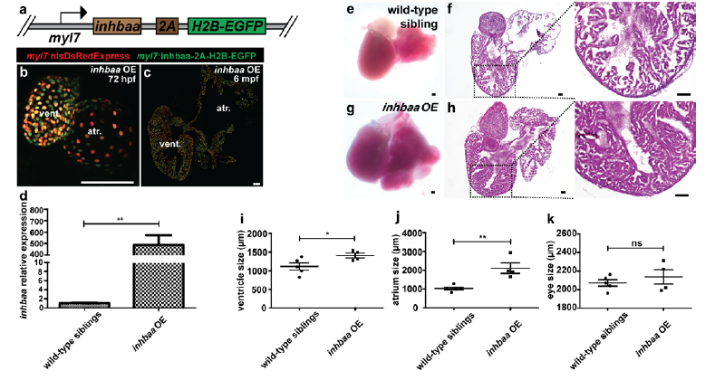
Generation and validation of inhbaa GOF line. (a) Schematic representation of CM-specific inhbaa OE transgene, Tg(myl7:inhbaa-2A-H2B-EGFP). (b) Heart of 72 hpf inhbaa OE larva in Tg(myl7:nlsDsRedExpress) background (native fluorescence). (c) Section of inhbaa OE adult fish heart in Tg(myl7:nlsDsRedExpress) background; α-DsRed (red), α-GFP (green). (d) RT–qPCR for inhbaa expression analysis in wild-type sibling and inhbaa OE adult hearts (n=2 x 3 cardiac ventricles assessed as 2 biological and 2 technical replicates). (e-h) Wild-type sibling and inhbaa OE adult hearts, H&E staining of heart sections and higher magnifications of H&E staining showing representative enlarged heart with dense trabeculae in inhbaa OE animals. (i-k) Quantification of ventricle, atrium and eye size in wild-type sibling (n=5) and inhbaa OE (n=4) adults. (data are mean ± s.e.m., ns: no significant changes observed, *P ≤ 0.05 and **P ≤ 0.0 by Student’s t-test, two-tailed). Scale bars, 100 μm. vent., ventricle; atr., atrium.