
|
Fig. 3
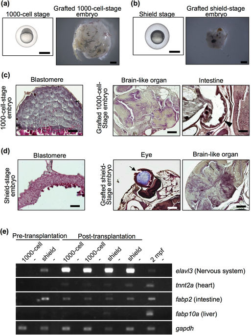
Failure of organ formation in grafted blastular and gastrular embryos. (a,b) Representative 1000-cell-stage (3 hpf) embryos (a), shield-stage (6 hpf) embryos (b) and grafted embryos two months after transplantation. Both embryo stages were subcutaneously transplanted into rag1 mutants. Scale bar: 1 mm. (c,d) Histology of 1000-cell-stage embryos (c) and shield-stage embryos (d) before and after transplantation. Sections were stained with haematoxylin and eosin. Although brain-like organs with an irregular shape and cell body arrangement were observed in all of the grafted early-stage embryos, other organs were not observed, except an eye (arrow) and intestine (arrow head) in a few grafted embryos. Scale bars: 200 µm (brain-like organ and eye), 100 µm (intestine), 50 µm (blastula and gastrula). (e) RT-PCR analysis of organ-specific genes in the grafted blastular and gastrular embryos at 2 months post-transplantation. Each experiment was conducted using two independent samples. Wild-type zebrafish at 2 mpf were used as positive controls. Negative controls lacking reverse transcriptase are located in alternating lanes (−). gapdh was used as a positive control.