
IMAGE
Fig. 5
- ID
- ZDB-IMAGE-180418-16
- Genes
- Publication
- Sarmah et al., 2017 - Embryonic Ethanol Exposure Affects Early- and Late-Added Cardiac Precursors and Produces Long-Lasting Heart Chamber Defects in Zebrafish
- All Figures
- Figures for Sarmah et al., 2017
Image
|
Figure Caption
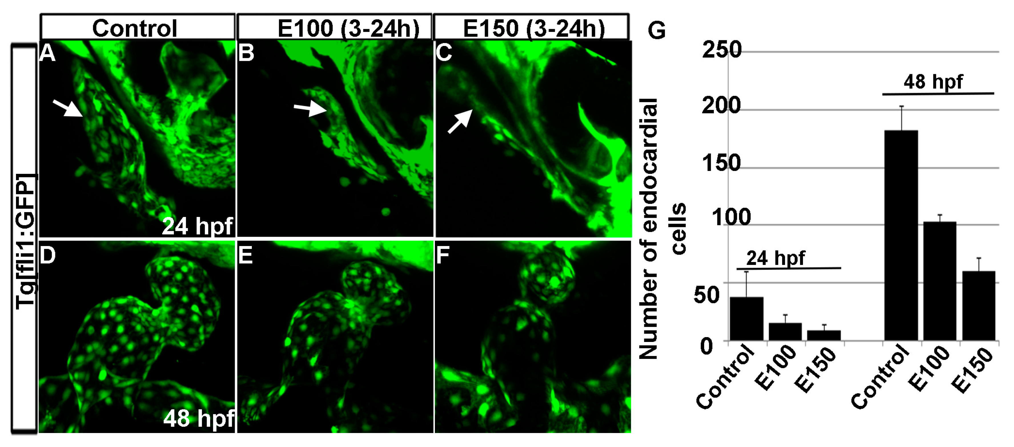
Fig. 5 Ethanol exposure reduced endocardial cell numbers. (A–C) Tg(fli1:EGFP) embryos showed endocardial lining in the linear heart tube in control embryo (A) and in ethanol-exposed embryos (B,C) at 24 hpf; (D–F) Tg(fli1:EGFP) embryos showed normal-shaped endocardium in the control embryo (D) and deformed endocardium with fewer endocardial cells in ethanol-exposed embryos at 48 hpf (E,F); (G) Graph shows the quantification of the endocardial cells at 24 and 48 hpf. White arrow: endocardial lining.
Figure Data
Acknowledgments
This image is the copyrighted work of the attributed author or publisher, and
ZFIN has permission only to display this image to its users.
Additional permissions should be obtained from the applicable author or publisher of the image.
Full text @ Toxics