
|
Fig. 1-S1
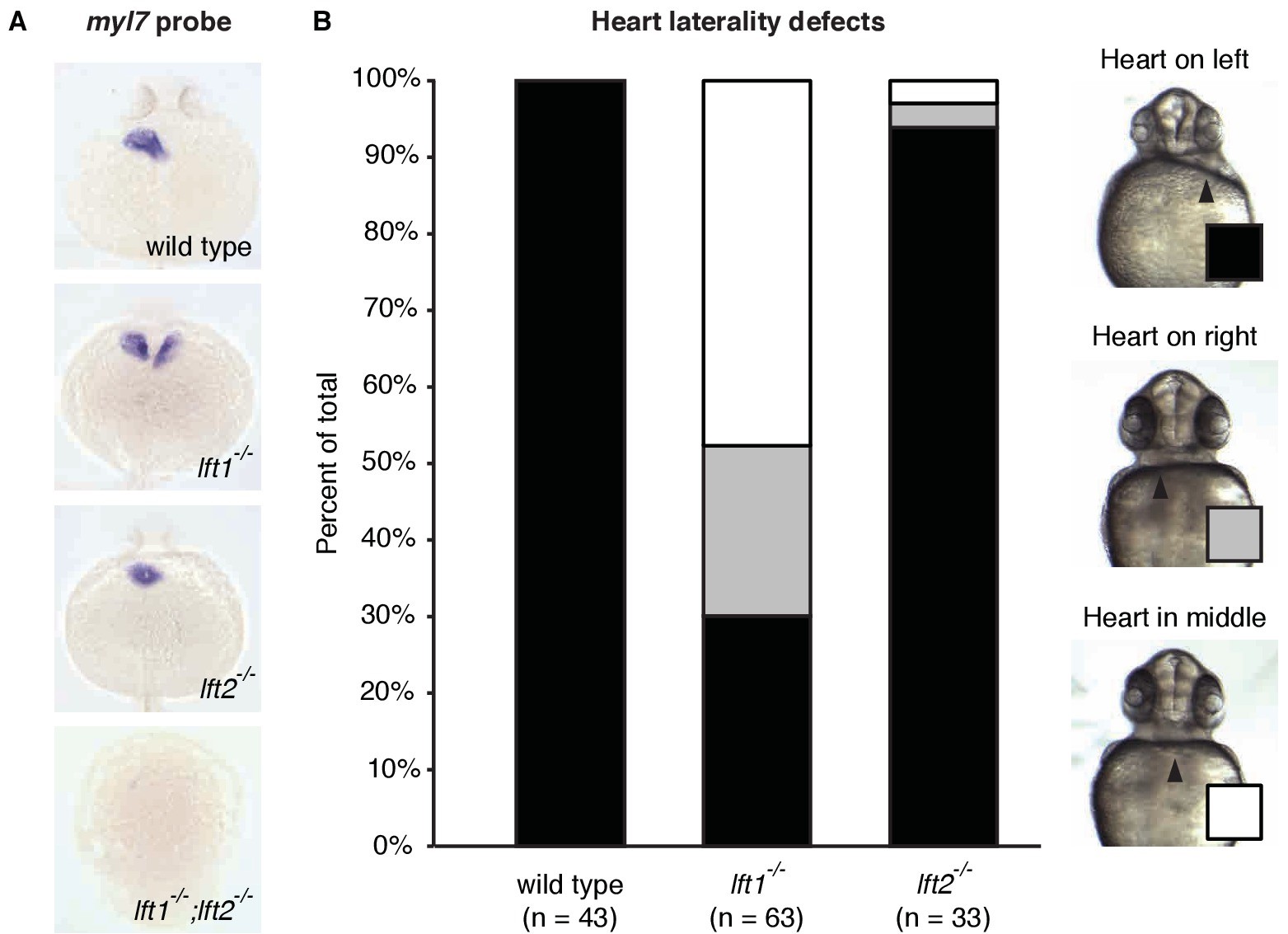
lefty1-/- mutants exhibit partially penetrant heart laterality defects.
(A) In situ hybridization using a probe against the heart muscle marker myl7 in 24 hr post-fertilization (hpf) wild type and lft mutant embryos (dorsal views). Most wild type and lft2-/- mutants exhibit myl7 expression on the left side. In contrast, some lft1-/- mutants exhibit bilateral myl7 expression, and lft1-/-;lft2-/- mutants express very little myl7 and typically fail to generate hearts (dorsal views). (B) Quantification of heart laterality defects in lft1-/- and lft2-/- mutants (ventral views). Live embryos were scored at 30 hpf. Whereas lft2-/- mutants typically exhibit normal heart laterality, lft1-/- mutants frequently have misplaced hearts. Despite this heart laterality defect, lft1-/- mutants are viable.