
Fig. S7
- ID
- ZDB-IMAGE-180410-34
- Publication
- Sawamiphak et al., 2017 - Transient cardiomyocyte fusion regulates cardiac development in zebrafish
- All Figures
- Figures for Sawamiphak et al., 2017
|
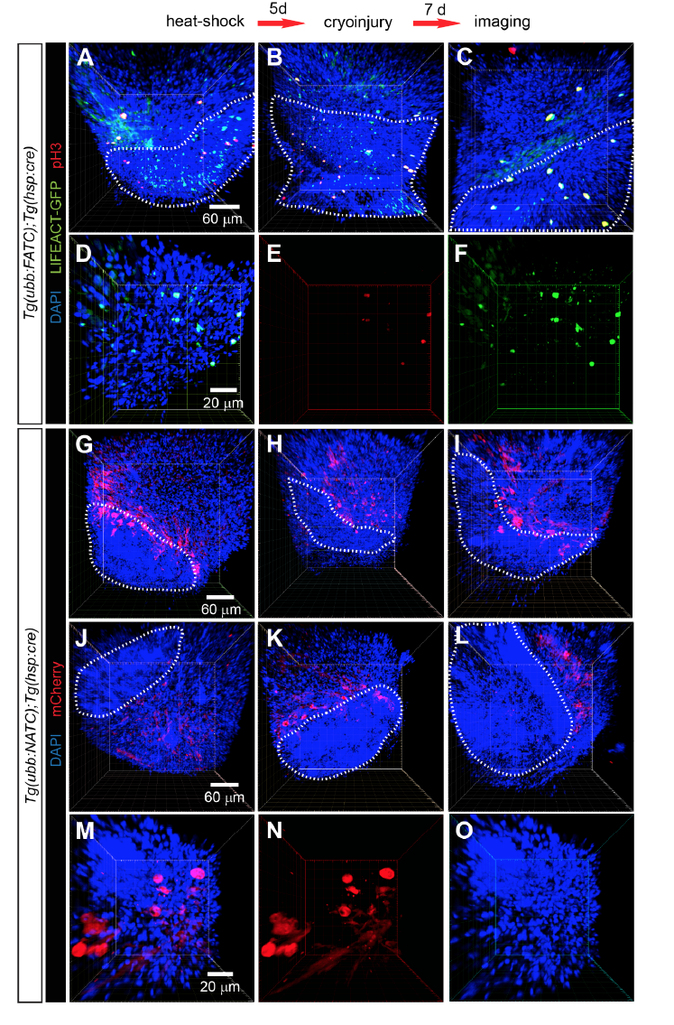
Fig. S7
F/NATC reporter expression correlates with cell proliferation during adult heart regeneration. A-F, FATC+ cells proliferate during cardiac regeneration. Tg(ubb:FATC);Tg(hsp:cre) fish (20 months old) were heat-shocked to induce cre expression, followed by cardiac cryoinjury 5 days later. Activation of FATC reporter, visualized by LIFEACTGFP expression (green), and mitotic activity, detected with phospho-Histone H3 immunostaining (pH3, red), in injured hearts was assessed at 7 dpi. Disorganized cells, visualized by DAPI (blue) staining, define the damaged area (white dashed lines). G-L, NATC-activated cells localized mainly adjacent to the damaged area. 12-month-old Tg(ubb:NATC);Tg(hsp:cre) fish were heatshocked to induce cre expression 5 days prior to cardiac cryoinjury. NTR-mCherry expression (red) was visualized by immunostaining at 7 dpi. DAPI (blue) nuclear staining shows disorganized cardiac cells in the injured area, outlined by white dashed lines. M-O, NTR-mCherry+ cells exhibit an atypical morphology that resembles immature/dedifferentiated cardiomyocytes. Each image is from an individual heart shown as a 3D volume rendering of a 100-400 μm thick confocal stack. 3 Tg(ubb:FATC);Tg(hsp:cre) and 6 Tg(ubb:NATC);Tg(hsp:cre) animals were examined.