
|
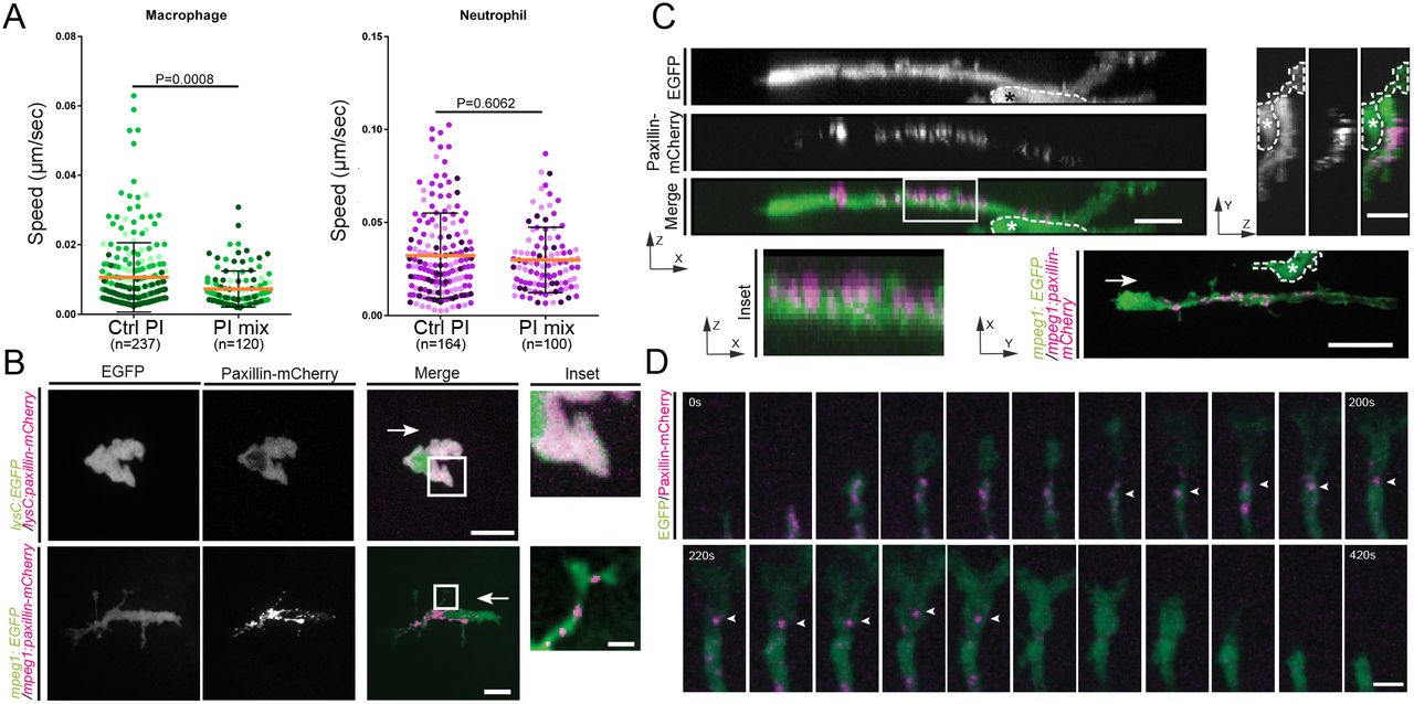
Fig. 2
Macrophages but not neutrophils require proteases for migration and form paxillin puncta. (A) Macrophage random migration is affected when a broad-range protease inhibitor mix is included in the larval medium. Under the same conditions neutrophil migration is not affected. The mean±s.d. speed is shown; P-values were calculated with a least squares means analysis. (B) Transiently expressed paxillin–mCherry forms puncta in macrophages, but has a cytoplasmic distribution in neutrophils. The inset column shows a magnification of the boxed region. Scale bar: 10 µm (2.5 µm for inset). See Movie 2. (C) Paxillin–mCherry puncta are located close to the membrane (zx view and inset) on only one side of the cell (zx and yz projections). Scale bars: 10 µm (z-projections); 20 µm (xy-projection). Asterisks mark a nearby cell not labeled with paxillin. See Movie 3. White arrows in B and C mark the direction of cell movement. (D) Dynamics of paxillin puncta inside macrophage protrusions (20 s intervals). Paxillin puncta disperse before retraction of the protrusion. Arrowheads mark the same paxillin punctum. Scale bar: 2.5 µm. Data are representative of seven independent experiments where all macrophages showed paxillin in puncta (a total of 27/27).