
|
Fig. s1
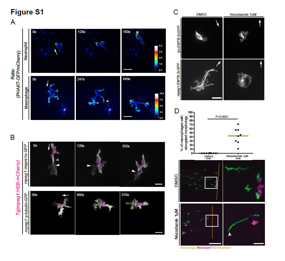
PHAKT-EGFP and microtubule localization during neutrophil and macrophage migration in live zebrafish. (A) Ratiometric imaging of neutrophils and macrophages. Neutrophils show presence of PIP3 at the leading edge. Macrophages show presence of PIP3 in all protrusions including the ones extending and retracting. Data are representative of 2 independent experiments for neutrophils and 3 independent experiments for macrophages. Scale bar, 20μm. Ratiometric scale shows numerical values. See Video 4. Asterisks mark non-specific signal. (B) Time-lapse imaging of macrophages transiently expressing Mapre1a-EGFP in a Tg(mpeg1:H2BmCherry) line. The signal is located at the front and back of the cell (white arrowhead show microtubules at the front) Scale bar, 20μm. Time-lapse images of transiently expressed gTubulin-EGFP in a Tg(mpeg1:H2B-mCherry) line. This probe shows the MTOC (green arrowhead) in the front of the nucleus in the direction of migration. White arrows indicate direction of migration. Scale bar, 20μm. (C) EMTB-3xGFP transiently expressed in neutrophils (lyz) and macrophages (mpeg1) showed that most microtubules are lost when nocodazole 1μM is present in the media. Scale bar, 20μm. Images are representative of 1 experiment. (D) Quantification of macrophages presenting a rear detachment phenotype, as a percentage of the total of macrophages during the timelapse movie. Nocodazole 1μM induced macrophage but not neutrophil elongation towards wounded tissues. White arrowhead labels trailing edge of an elongated macrophage. Scale bar, 100μm and 25μm for insets. See Video 7. Data are representative of 2 blind tests, from 4 independent experiments, with two-tailed unpaired t test.