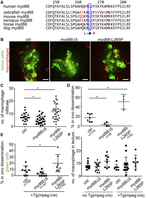
Fig. 5 Altering Macrophage Recruitment to the Tumor by Altering Myd88 Function Affects Tumor Cell Dissemination (A) Nucleotide alignment of vertebrate Myd88 sequences flanking conserved amino acid 265 (blue box), which is mutated to P to make a constitutively active form of the protein. (B) Macrophages (red) marked by Tg(mpeg1:tdTomato)w201 infiltrating the xenotransplanted tumor cells (green) in control (left, n = 5 experiments), myd88w187 mutant (middle, n = 7 experiments), and Tg(mpeg:myd88-L265P)fh507 transgenic larva (right, n = 4 experiments). Scale bars, 30 μm. (C and D) Quantification of macrophage infiltrate (C) and in vivo dissemination (D) after xenotransplantation into control, myd88w187 mutant, and Tg(mpeg:myd88-L265P)fh507 transgenic larva. (C) One-way ANOVA (p < 0.0001), followed by non-parametric unpaired t test (p < 0.05 [between ctrl and myd88w187], p < 0.05 [between ctrl and myd88-L265P]). Each data point is a larva. Error bars are mean ± SD. (D) One-way ANOVA (p < 0.0001), followed by non-parametric unpaired t test (p = 0.04 [between ctrl and myd88w187], p = 0.01 [between ctrl and myd88-L265P]). Error bars are mean ± SD. For (C) and (D), asterisks indicate p < 0.05. (E) Quantification of the percent larvae with GFP+ disseminated tumor cells after transplantation into mosaically expressing mpeg1:Cre animals in a control, myd88w187 mutant, or Tg(mpeg:myd88-L265P)fh507 background. One-way ANOVA (p < 0.0001), followed by non-parametric unpaired t test, p < 0.05 (between ctrl and myd88-L265P). Error bars are mean ± SD. For (D) and (E), each data point is an independent experiment and n = number of larvae. Asterisk indicates p < 0.05. (F) Each data point represents the number of macrophages in a tectum of a larva, as determined by single time point z stacks (80 μm) in the tectum. One-way ANOVA, p = 0.8, not significant.
Reprinted from Developmental Cell, 43, Roh-Johnson, M., Shah, A.N., Stonick, J.A., Poudel, K.R., Kargl, J., Yang, G.H., di Martino, J., Hernandez, R.E., Gast, C.E., Zarour, L.R., Antoku, S., Houghton, A.M., Bravo-Cordero, J.J., Wong, M.H., Condeelis, J., Moens, C.B., Macrophage-Dependent Cytoplasmic Transfer during Melanoma Invasion In Vivo, 549-562.e6, Copyright (2017) with permission from Elsevier. Full text @ Dev. Cell

