
Image
|
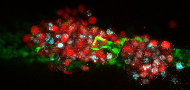
Figure Caption
Fig. 6 Confocal image of immunohistochemistry with Ki67 in a whole 6 dpi (fli:GFP) Casper zebrafish embryo. There were 200 to 400 CRMM-1 td-Tomato CM cells injected into the duct of Cuvier. We see tumor cell (red) migration outside the vessels (green); cell proliferation is indicated by Ki67 staining (blue). This image of the tail of a live embryo was acquired by confocal microscope (×20 dry objective). Similar images were obtained from all three CM cell lines in >10 independent experiments.
Acknowledgments
This image is the copyrighted work of the attributed author or publisher, and
ZFIN has permission only to display this image to its users.
Additional permissions should be obtained from the applicable author or publisher of the image.
Full text @ Invest. Ophthalmol. Vis. Sci.