
|
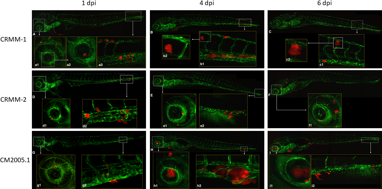
Fig. 4 Confocal micrographs of the observed phenotypes at 1, 4, and 6 dpi after engraftment of three CM cell lines via the duct of Cuvier in (fli:GFP) Casper zebrafish embryos. At 1 dpi, CRMM-1 (A), CRMM-2 (D), and CM2005.1 (G) cells were already inside the eye (a1, a2, d1, g1) and in the tail (a3, d2, g2). At 4 (B, E, H), and 6 dpi (C, F, I), cells formed clusters in the tail and in the eye in all three cell lines (data not shown). The clusters were more evident in the tail (h2) and in the eye (h1, i1) after injection of cell line CM2005.1. The three cell lines (data not show) grew inside (a3, b1, d2, e2, g2, h2, i2), outside (b2), and around (c2) the vessels and the cells could be found inside the eye (f1, i1) until 6 dpi. The images were acquired using a Leica TCS SPE confocal microscope and managed in ImageJ software. Images (A–I) ×10 dry objective. All the other images: ×20 dry objective. Red: cells labeled with tdTomato; green: GFP-endothelial cells of the (fli:GFP) Casper lines.