
|
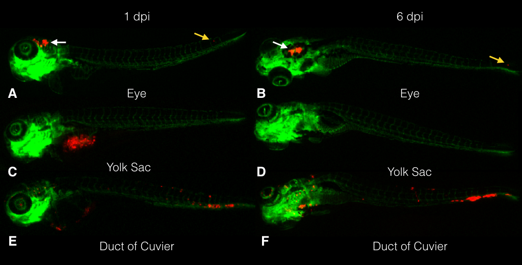
Fig. 1 Stereo fluorescence image of zebrafish embryos engrafted with CM cells (vasculature in green and CM cells in red). The embryos were injected at 2 dpf with CRMM-1 CM cells labeled with tomato-red (red). Photographs taken of the same embryo that had been injected with CM cells around the eye at 1 (A) and 6 dpi (B), showing cells inside the head (white arrows) and in the tail (yellow arrow). Following injection in the yolk sac, an embryo shows the cells in the yolk sac at 1 (C), but not 6 dpi (D). After injection of cells into the duct of Cuvier, cells are seen inside the circulation at 1 dpi (E), mainly in the tail and inside the eye. The same embryo shows a cluster in the tail and cells inside the eye at 6 dpi (F). The stereo fluorescent images (original magnification: ×20) are representative of >10 independent experiments.