
|
Fig. 5
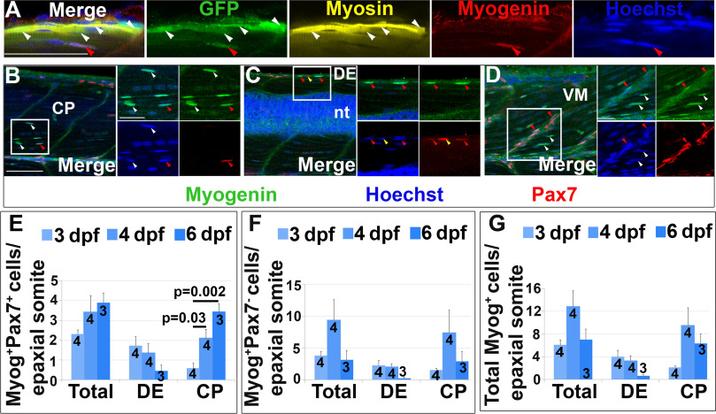
Pax7+ cells differentiate in specific somite regions. Single confocal planes of 4 dpf wholemount immunofluorescence in lateral view. Scale bars 50 µm. A. pax7a:GFP;pfe/pfe larva showing GFP in a MyHC+ muscle fibre (white arrowheads). A deep GFP+MyHC- cell co-labels with Myogenin (red arrowheads). B-D. Pax7 and Myogenin in epaxial somite of wt larva showing Pax7+Myog+ cells (red arrowheads) in CP (B), DE (C) and VM (D), Pax7+Myog- cells (yellow arrowheads) in DE (C) and Pax7-Myog+ cells (white arrowheads) in CP (B,D). Note the reduced Pax7 and Myog signal in Pax7+Myog+ cells. E-G. Time course and location of Myog+Pax7+ (E), Myog+Pax7- (F) and total Myog+ (G) cells.
Reprinted from Developmental Biology, 431(2), Roy, S.D., Williams, V.C., Pipalia, T.G., Li, K., Hammond, C.L., Knappe, S., Knight, R.D., Hughes, S.M., Myotome adaptability confers developmental robustness to somitic myogenesis in response to fibre number alteration, 321-335, Copyright (2017) with permission from Elsevier. Full text @ Dev. Biol.