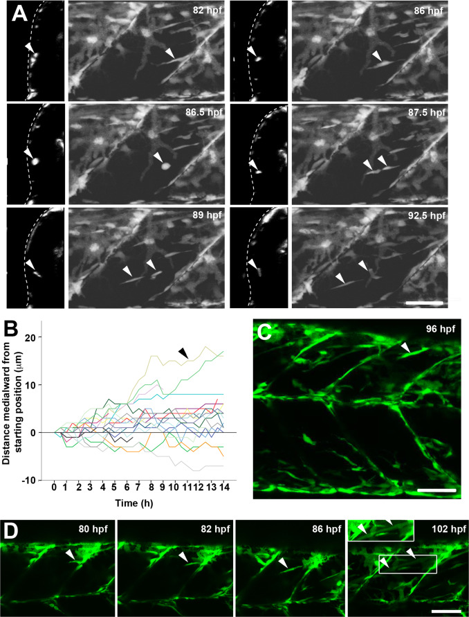
IMAGE

Fig. 4

ID
ZDB-IMAGE-180322-26
Source
Figures for Roy et al., 2017
Image
Figure Caption

Fig. 4

ax7a:GFP+ cells migrate into deep central myotome and differentiate. Confocal maximum intensity Z projections and orthogonal YZ views of live pax7a:GFP;pfe/pfe larvae in lateral view. A. Timelapse of 3.5–4 dpf larva taken every 30 min for 14 h showing migration of a cell from the posterior vertical myoseptum into the deep myotome (arrowheads). Note rounding up, division and separation of daughters. B. Analysis of distance moved towards midline in Z plane for each cell in the epaxial somite from the timelapse shown in A. Note that total movement of cells is often greater, as migration in the anteroposterior and dorsoventral planes (XY) is not shown. Each cell was measured relative to its starting position. Arrowhead indicates the cell highlighted in A. C. A cell extending from the dorsal edge into the deep myotome (arrowhead). D. Timelapse of myotube formation from a cell entering the myotome from the posterior VM (arrowheads). Between 86 and 102 hpf the GFP became diluted in the extended fibre (box is shown with contrast enhancement in inset above). Bars 50 µm.

Acknowledgments
This image is the copyrighted work of the attributed author or publisher, and ZFIN has permission only to display this image to its users. Additional permissions should be obtained from the applicable author or publisher of the image.

Reprinted from Developmental Biology, 431(2), Roy, S.D., Williams, V.C., Pipalia, T.G., Li, K., Hammond, C.L., Knappe, S., Knight, R.D., Hughes, S.M., Myotome adaptability confers developmental robustness to somitic myogenesis in response to fibre number alteration, 321-335, Copyright (2017) with permission from Elsevier. Full text @ Dev. Biol.