
|
Fig. 3
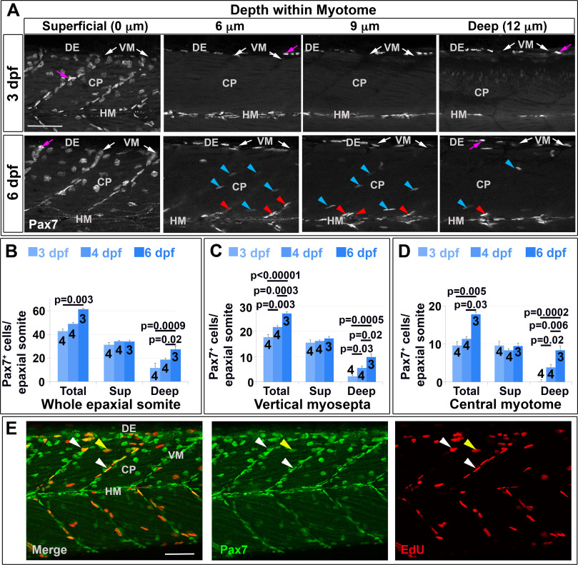
Pax7+ nuclei increase in deep myotome. Pax7 (A-E) and EdU (E) labelling in wholemount wt larvae. Single confocal slices of zebrafish larvae in lateral view. A: Flattened dehydrated embryos imaged at the indicated depths (full larval thickness approximately 35 µm). At 3 dpf, MPCs accumulate superficially near VM (white arrows) but are absent deeper within myotome. Xanthophores (purple arrows) are rare and bright. By 6 dpf, Pax7+ nuclei appear deep at the VM (red arrowheads) and CP (blue arrowheads). B-D: Numbers of Pax7+ nuclei in epaxial somites 16–18 of whole mount larvae increase with age. Mean±S.E.M. The small error bars indicate tight regulation of Pax7+ cell numbers. Number of embryos scored is indicated within the columns. E: Co-localization of Pax7 and EdU in MPCs of 4 dpf larva both at VM (white arrowheads) and CP (yellow arrowheads). Vertical myoseptum (VM), central portion (CP), horizontal myoseptum (HM), dorsal edge (DE). Bars 50 µm.
Reprinted from Developmental Biology, 431(2), Roy, S.D., Williams, V.C., Pipalia, T.G., Li, K., Hammond, C.L., Knappe, S., Knight, R.D., Hughes, S.M., Myotome adaptability confers developmental robustness to somitic myogenesis in response to fibre number alteration, 321-335, Copyright (2017) with permission from Elsevier. Full text @ Dev. Biol.