
Fig. s2
- ID
- ZDB-IMAGE-180322-11
- Publication
- Mojib et al., 2017 - Zebrafish aversive taste co-receptor is expressed in both chemo- and mechanosensory cells and plays a role in lateral line development
- All Figures
- Figures for Mojib et al., 2017
|
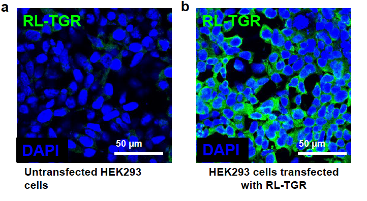
Fig. s2 Specificity of custom-generated anti-RLTGR antibody with RL-TGR protein. (a) Confocal image of the untransfected Human Embryonic Kidney (HEK) 293 cells immunostained with anti-RL-TGR antibody (green) followed by staining with DAPI that stains nuclei blue. (b) Confocal image of HEK293 cells transfected with mammalian expression vector, pcDNA3.1 (+) containing coding region of rltgr and then immunostained with anti-RL-TGR antibody (green) followed by staining with DAPI (blue). The green fluorescence observed in cells expressing recombinant RL-TGR protein indicates that the custom generated antibodies recognize specifically the RL-TGR protein.