
Fig. 6
- ID
- ZDB-IMAGE-180321-17
- Publication
- Campbell et al., 2017 - Phosphodiesterase Inhibitors Sildenafil and Vardenafil Reduce Zebrafish Rod Photoreceptor Outer Segment Shedding
- All Figures
- Figures for Campbell et al., 2017
|
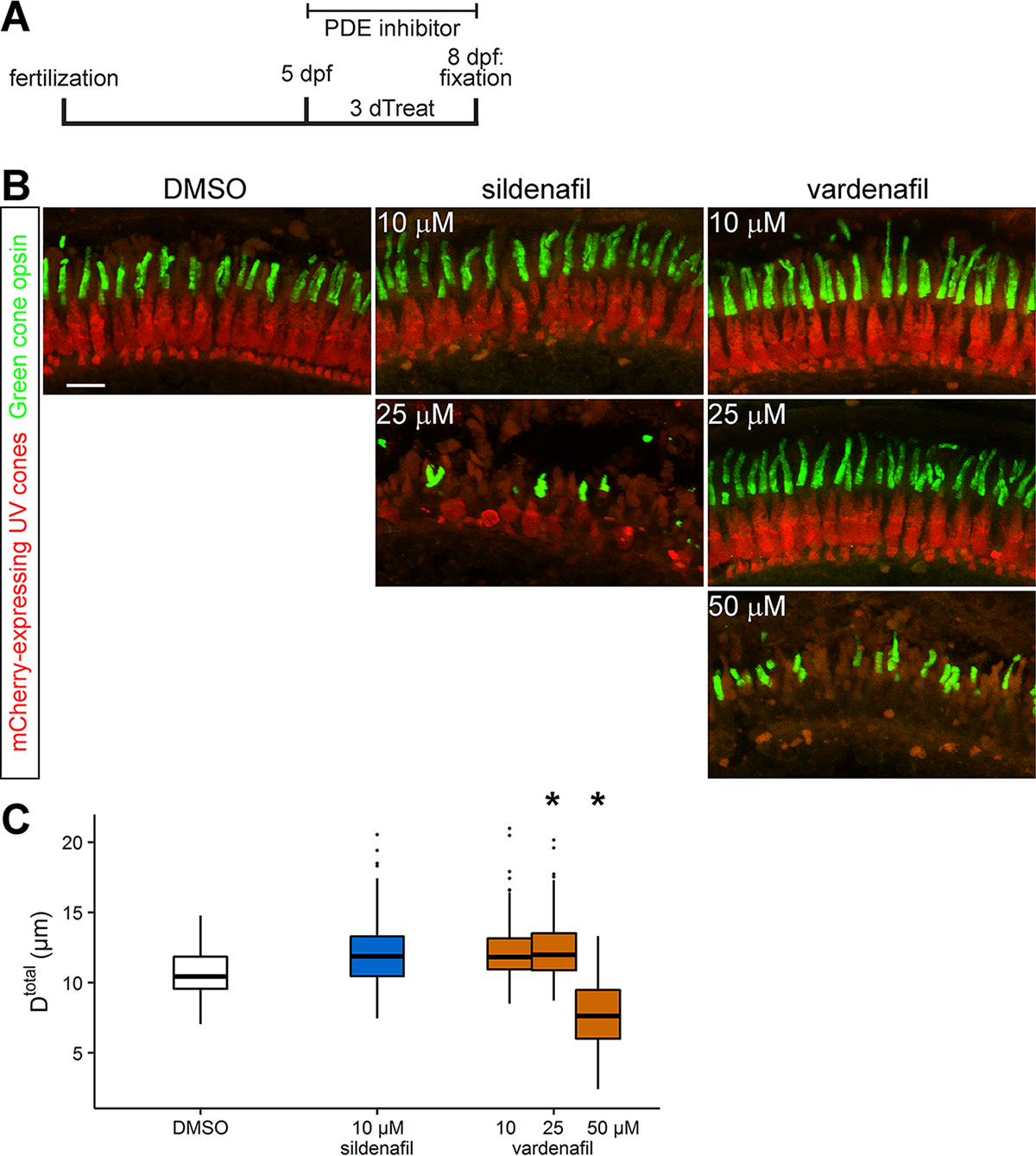
Fig. 6 Cone photoreceptors are sensitive to sildenafil and vardenafil. (A) Schematic timeline for examining green cone outer segment length in 8 dpf Tg(SWS1:mCherry); albb4/b4 larvae after bathing in phosphodiesterase inhibitor for 3 days (dTreat). (B) Representative images of photoreceptor layers from larvae treated with 0.05% DMSO, sildenafil, and vardenafil at indicated concentrations. Retinal sections were immunolabeled with green cone opsin antibody (green); ultraviolet cones express mCherry (red). Images are projections of a subset of z-sections totaling 7.33 μm. Scale bar: 10 μm. (C) Quantification of green cone outer segment full length (Dtotal). Lower and upper hinges of box correspond to first and third quartiles; middle corresponds to median; whiskers extend 1.5 * interquartile range above and below the hinges; dots represent outliers. Graph represents n = 5 fish/condition (DMSO = 162 outer segments, sildenafil = 169 outer segments, and vardenafil ≥ 175 outer segments). *95% CI of difference compared to control does not span zero.