
|
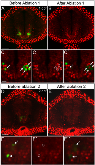
Fig. 3
Before ablation 1 and 2, Pth4:eGFP+/Bactin2:H2A-mCherry+ transgenic embryo shows two bilateral clusters of neurons in the hypothalamic area (A and D). After several rounds of ablation the eGFP-expressing cells are completely eliminated (B and E). Ablation processes example (C-C´´and F-F´´): (C and F) arrows indicate eGFP-expressing cells chosen for ablation; nuclear region of interest for targeted elimination of Pth4:eGFP neurons is defined by circles, based on red fluorescence from nuclear marker Bactin2 (C´ and F´); immediately after ablation Pth4:eGFP neurons are eliminated (marked by arrows in C´´ and F´´). Note that the untargeted cells around the eGFP-expressing cells ablated are intact (C´´ and F´´). A confocal z-stack projection (A, B, D and E) or single 2 μm thick z-plane slice (C-C´´ and F-F´´) are shown. Scales bars: 50 μm (A, B, D and E); 15 μm (C-C´´ and F-F´´).