
|
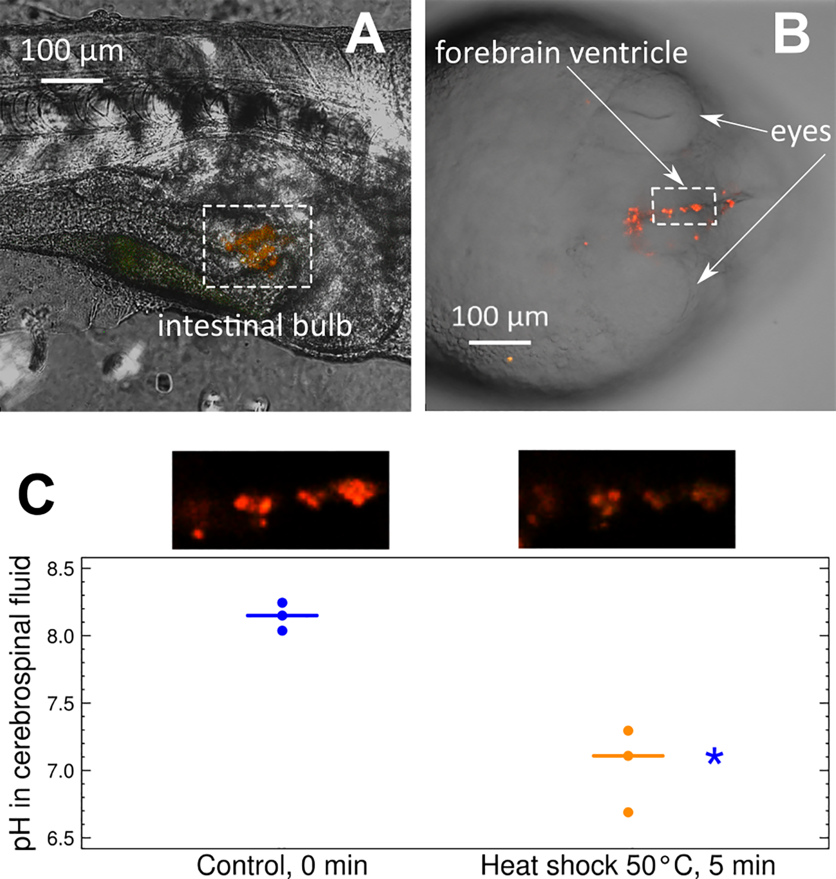
Fig. 2 Visualization of MBMs and pH measurements in zebrafish embryos.
(a) Images of MBMs in the intestine of a zebrafish embryo, combined green (587 nm) and red (627 nm) channels. (b) Images of MBMs in brain ventricle of a zebrafish embryo, combined green (587 nm) and red (627 nm) channels. (c) pH in cerebrospinal fluid monitored by MBMs under control and heat shock conditions with respective original images of MBMs in the brain ventricle of the same individual. Blue indicates control conditions; orange indicates heat shock exposure. * indicates a statistically significant difference from the control with p-value < 0.05.