Fig. 5
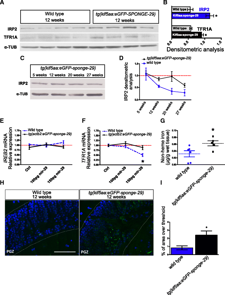
Genetic repression of miR-29 chronically affects iron homeostasis. a, b Western blot of IRP2 and TFR1A in 12-week-old tg(kif5aa:eGFP-sponge-29) (n = 4) and wild-type (n = 4) fish brain extracts and (b) relative densitometric analysis (*P < 0.05; **P < 0.01, Mann–Whitney U-test). c Representative Western blot of IRP2 in brain extracts of kif5a:sponge-29 at different ages (5, 12, 20, 27). In a–c α-TUBULIN was used as loading control. d Comparison of densitometric analysis calculated on Additional file 8a and b. The blue line represents IRP2 expression in tg(kif5aa:eGFP-sponge-29) animals, gray line represents expression in wild-type fish. Values were normalized to the mean of 5 weeks value (Kruskal–Wallis test, P = 0.0939, n = 3 animals for each age point). e, f Expression level at 24 hpf of Ireb2 and Tfr1a upon miR-29 mimics injection. The expression level was determined by RT-qPCR. Statistical significance was assessed by one-way ANOVA with post-hoc Tukey’s test (*P < 0.05), the analysis was performed on total RNA extraction from 30–40 embryos for each condition. Error bars indicate standard errors of means. g Brain non-heme iron content (μg/g wet tissue) in kif5a:eGFP-sponge-29 (n = 5) compared to wild-type (n = 6) at age 12 weeks (*P < 0.05, Mann–Whitney U-test). h Representative images of lipofuscin accumulation in the optic tectum of 12-week-old kif5a:eGFP-sponge-29 and wild-type fish brains. Lipofuscin auto-fluorescent granules (green) were detected with ApoTome microscope, counterstained with DAPI (blue). Scale bar: 50 μm. i Quantification of lipofuscin density based on percentage of area over threshold, n = 6 (*P < 0.05; Mann–Whitney U-test)

