
|
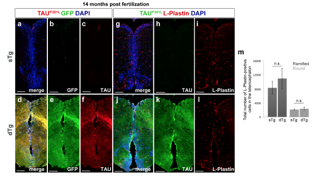
Fig. s11 (a) Immunohistochemistry (IHC) for TAUP301L (red) and GFP (green) on coronal sections of telencephalon of a 14-month old sTg animal. (b,c) Individual fluorescent channels for TAUP301L (b) and GFP (c). (d) IHC for TAUP301L and GFP on coronal sections of telencephalon of a 14 month-old dTg animal. (e,f) Individual fluorescent channels for TAUP301L (e) and GFP (f). (g) IHC for L-Plastin and TAUP301L on coronal sections of telencephalon of a 14 month-old sTg animal. (h,i) Individual fluorescent channels for TAUP301L (h) and L-Plastin (i). (j) IHC for L-Plastin and TAUP301L on coronal sections of telencephalon of a 14-month old dTg animal. (k,l) Individual fluorescent channels for TAUP301L (k) and L-Plastin (l). (m) Quantification of round and ramified L-Plastin-positive cells in the telencephalon of 14-month-old sTg and dTg animals. Values represent mean ± s.e.m. *: p<0.05, **: p<0.01, ***: p<0.005. Scale bars equal 50 μm. n = 5 fish and >25 histological sections for every staining.