
|
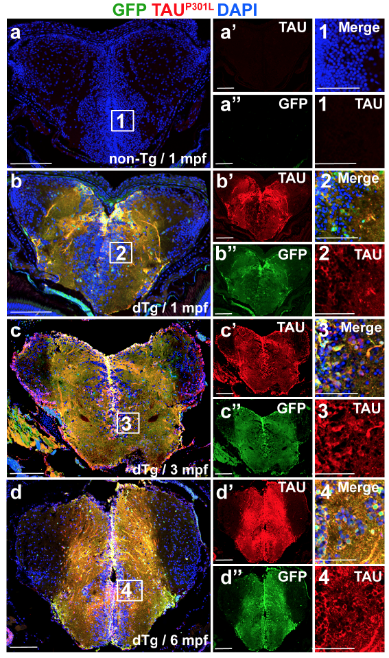
Fig. s3 (a) Immunohistochemistry (IHC) for TAUP301L (red) and GFP (green) on coronal sections of telencephalon of a 1 month-old nontransgenic animal. (a’, a’’) Individual fluorescent channels for TAUP301L (a’) and GFP (a’’). (1) The enlarged view of the inset in a. (b) IHC for TAUP301L and GFP on coronal sections of telencephalon of a 1-month old dTg animal. (b’, b’’) Individual fluorescent channels for TAUP301L (b’) and GFP (b’’). (2) The enlarged view of the inset in b. (c) IHC for TAUP301L and GFP on coronal sections of telencephalon of a 3-month old dTg animal. (c’, c’’) Individual fluorescent channels for TAUP301L (c’) and GFP (c’’). (3) The enlarged view of the inset in c. (d) IHC for TAUP301L and GFP on coronal sections of telencephalon of a 6-month old dTg animal. (d’, d’’) Individual fluorescent channels for TAUP301L (d’) and GFP (d’’). (4) The enlarged view of the inset in d. Scale bars equal 50 μm. n = 4 fish and >25 histological sections for every staining.