Image
Figure Caption
Fig. 2
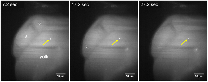
Figure 2: LSFM images of a static parasite in a 48 hpf larva. The T. cruzi parasite (yellow arrow) remains adhered to the walls of the yolk sac, throughout the time-lapse sequence (7.2 s, 17.2 s, and 27.2 s), about ~15 min after parasite injection. No change in position of the parasite is observed during an acquisition period of at least 30 s. a, Atrium; v, Ventricle. Scale bar = 50 µm.
Acknowledgments
This image is the copyrighted work of the attributed author or publisher, and
ZFIN has permission only to display this image to its users.
Additional permissions should be obtained from the applicable author or publisher of the image.
Full text @ J. Vis. Exp.

