
|
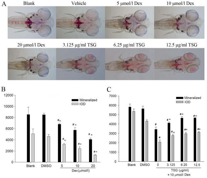
Fig. 5
Effects of Dex and TSG on zebrafish larvae at 9 days post fertilization. (A) Ventral view of Alizarin red whole-mount preparations (magnification, ×100). Areas of calcified matrix in craniofacial skeleton are stained red, while otoliths (not bony structures) appear brown/black. Compared with the blank and vehicle (0.5% DMSO) control groups, the groups treated with 5, 10 and 20 µmol/l Dex demonstrated marked decrease in area and density of red staining. The TSG-treated groups (3.125, 6.25 and 12.5 µg/ml) presented significantly increased mineralized area and density of red staining. (B) Effects of Dex on mineralized area and IOD of mean pixel number of zebrafish larvae. #P<0.05 vs. blank group; *P<0.05 vs. DMSO control group. (C) Effects of TSG on mineralized area and IOD of mean pixel number of zebrafish larvae. #P<0.05 vs. DMSO control group; *P<0.05 vs. 10 µmol/l Dex group. Dex, dexamethasone; TSG, tetrahydroxystilbene glucoside; IOD, integrated optical density; DMSO, dimethyl sulfoxide.