
|
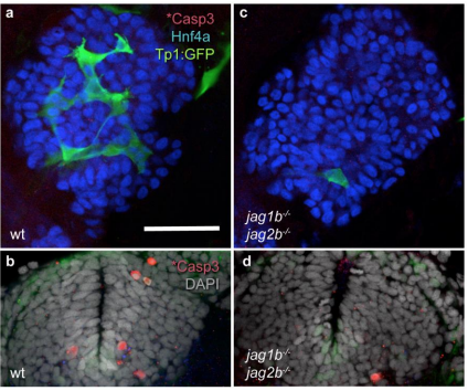
Fig. s5 Supplementary figure 5. Apoptosis does not contribute to the lack of intrahepatic duct cells in jag1b and jag2b double homozygous mutants. (a-d) Whole mount immunofluorescent images of 54 hpf sibling (a,b) and double mutant (c,d) embryos labeled for cleaved Caspase3 (*Casp3) expression. Sibling (a) and double mutant (c) livers labeled for Hnf4a (blue) show no signs of apoptosis based on a lack of cleaved Caspase3 (red) expression or blebs of Tp1:GFP+ (green), whereas the ventral forebrains (b,d, with DAPI channel also shown) of the same embryos (from a,c) show normal apoptosis patterns as indicated by variable presence of cleaved Caspase3 (red). Scale bar 50μM. Representative samples, n= 4 siblings, 4 jag1b-/-;jag2b-/-