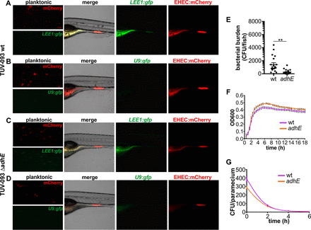
Fig. 5
LEE expression and colonization of EHEC ΔadhE. (A) EHEC TUV 93-0 wild type transformed with mCherry and LEE1::gfp grown planktonically in E3 medium and in zebrafish. (B) EHEC TUV 93-0 wild type transformed with mCherry and promoterless U9::gfp grown planktonically and in zebrafish. (C) EHEC TUV 93-0 isogenic ΔadhE mutant transformed with mCherry and LEE1::gfp grown planktonically and in zebrafish. (D) EHEC TUV 93-0 isogenic ΔadhE mutant transformed with mCherry and promoterless U9::gfp grown planktonically and in zebrafish. (E) Bacterial burden was determined by dilution plating on EHEC selective agar. Individual data points (n = 15 for each condition), means, and SD are shown. Statistical significance was determined using Student’s t test (**, P < 0.01). (F) Growth curves of TUV 93-0 wild type (pink) and ΔadhE mutant (orange). Means (n = 3) and SD are shown. (G) Degradation profiles for TUV 93-0 wild type (pink) and ΔadhE mutant (orange) in P. caudatum as determined by lysis of P. caudatum and dilution plating. Regression analysis showed there is no difference between the slopes (P = 0.2909).

